
Figure 5. sFRP3 inhibits EndMT.
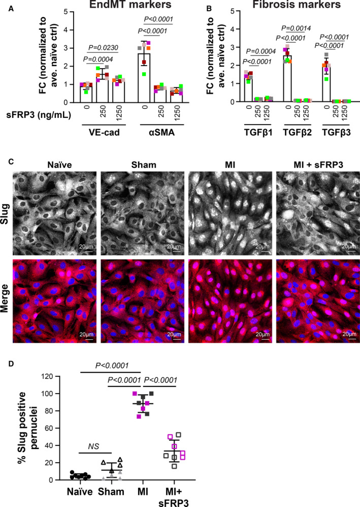
A and B, Mitral VECs were treated with MI plasma (n=6) supplemented with 0, 250, or 1250 ng/mL sFRP3 for 24 hours before qPCR. Mean±SD from three independent assays were graphed. P values were calculated using nonparametric Mann‐Whitney test. C, Mitral VECs were treated with post‐MI plasma±sFRP3 (250 ng/mL) for 24 hours before immunofluorescent staining using anti‐Slug (red). Naïve and sham plasma served as control. DAPI was used to stain nuclei (blue). Both black‐and‐white and colored merged images are shown to ease visualization (scale bar: 20 µm). D, Number of nuclei positive for Slug divided by total nuclei from 4 wells—duplicates incubated with 2 individual plasmas for each group of animals—were graphed. P values were calculated using 1‐way ANOVA with Sidak’s multiple comparisons test. EndMT indicates endothelial‐to‐mesenchymal transition; FC, fold changes; LVEF, left ventricular ejection fraction; MI, myocardial infarction; qPCR, quantitative polymerase chain reaction; sFRP3, secreted frizzled‐related protein 3; and VEC, valve endothelial cell.