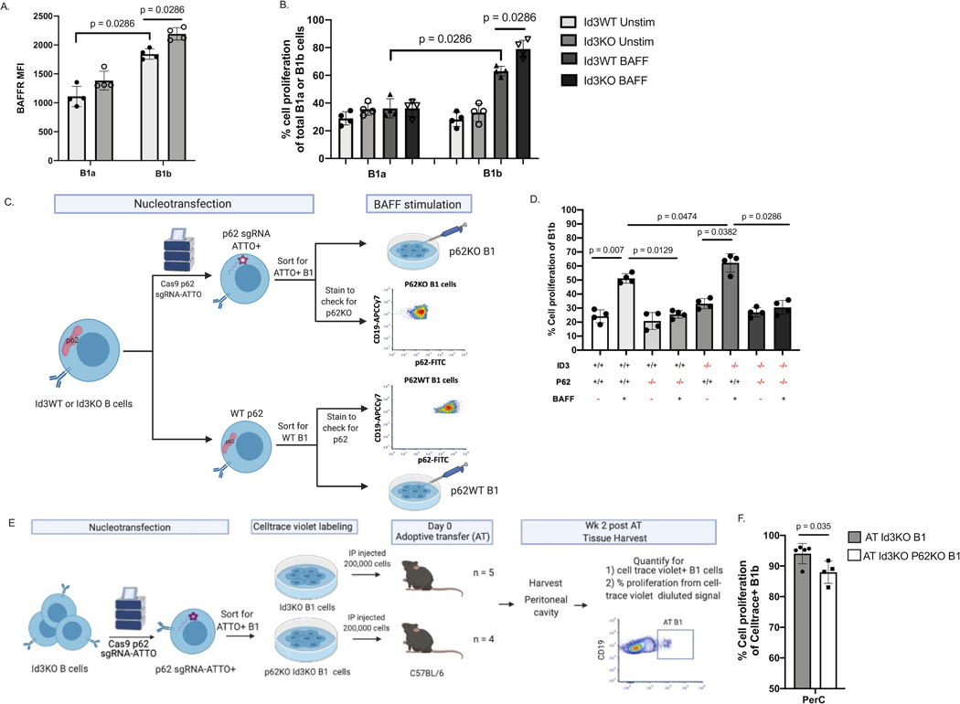
Figure 2: Loss of P62 significantly reduces B1b cell proliferation in vitro and in vivo.
(A) MFI of BAFFR in Id3WT and Id3KO B1a and B1b cells (n = 4 per each group). (B) percentage of cell proliferation measured by Celltrace-violet on Id3WT and Id3KO B1a and B1b cells under unstimulated and BAFF stimulated conditions (n = 4 per each group). (C) Schematics showing nucleofection of Cas9 p62 targeted gRNA labeled with ATTO fluorophore ribonucleoprotein complex (p62-gRNA-RNP) into murine Id3WT and Id3KO B cells following with sorting for ATTO+ B1 cells to select for p62KO B1 cells. (D) Percentage of cell proliferation of Id3WT and Id3KO B1b cells with conditional p62KO and BAFF stimulation measured by Celltrace-violet (n = 4 per each group). (E) Schematics showing nucleofection of p62-gRNA-RNP into Id3KO B cells and sorting for ATTO+ B1 cells to select for p62KO B1 cells, following with AT of Id3KOp62KO or Id3KO B1 cells into C57BL/6 to quantify proliferation of AT cells in various tissue compartment 2 wks post AT. (F) Percentage of cell proliferation of AT Id3KO (grey, n = 5) and Id3KOp62KO (white, n = 3) B1b cells 2 wk post transfer measured by Celltrace-violet. Results were represented with Mean ± SD with unpaired Mann-Whitney t-test.

