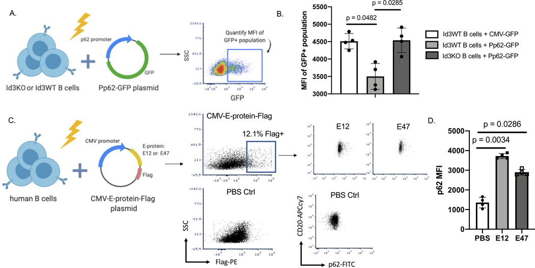
Figure 3: Loss of Id3 and increased expression of E12 and E47 promote p62 promoter activation.
(A) Schematics showing nucleofection of p62 promoter GFP plasmid (Pp62-GFP) or CMV-GFP plasmid control in murine Id3WT or Id3KO B cells to quantify GFP expression. (B) MFI of GFP+ B cells to compare GFP expression between Id3WT B cells transfected with CMV-GFP control (n = 4), Id3WT B cells transfected with Pp62-GFP (n = 4) and Id3KO B cells transfected with Pp62-GFP (n = 4). (C) Schematics demonstrating nucleo-transfection CMV-E12-Flag and CMV-E47-Flag plasmids into human B cells to measure P62 expression driven by overexpression of E-protein. (D) MFI of P62 compared across PBS control (n = 4), CMV-E12-Flag (n = 4) and CMV-E47-Flag (n = 4) transfection in human B cells. Results were represented with Mean ± SD. Statistical analyses were performed using Kruskal-Wallis test with Dunn’s correction for multiple comparison.

