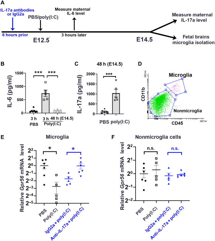
Fig. 1. MIA down-regulates microglial Gpr56 expression in fetal brains in an IL-17a–dependent manner.
(A) A schematic timeline for MIA induction. (B) Serum concentration of IL-6 was significantly up-regulated at 3 hours after poly(I:C) injection and went back to baseline 48 hours later at E14.5 (n = 5). (C) Serum concentration of IL-17a was significantly up-regulated at E14.5 48 hours after poly(I:C) injection (n = 5). (D) Fluorescence-activated cell sorting (FACS) strategy to collect CD11b+CD45medium microglia and CD11b−CD45− cells as nonmicroglial cells. (E) Relative levels of Gpr56 mRNA in microglia isolated from E14.5 fetal brains whose mothers were treated with PBS (n = 6), poly(I:C) (n = 5), IgG2a isotype control antibodies and poly(I:C) (n = 5), and IL-17a antibodies and poly(I:C) (n = 6). (F) Relative levels of Gpr56 mRNA in nonmicroglia cells isolated from E14.5 fetal brains. PBS versus poly(I:C): P = 0.55; IgG2a + poly(I:C) versus IL-17a antibodies + poly(I:C): P = 0.69. Unpaired Student’s t test. n.s., not significant. *P < 0.05 and ***P < 0.001. Data presented as means ± SEM.

