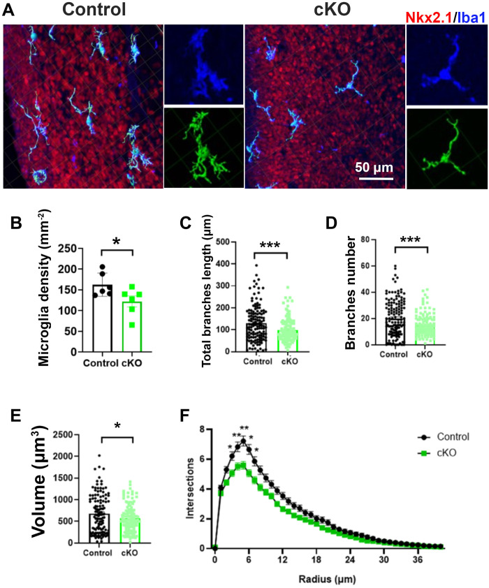
Fig. 5. Deleting microglial Gpr56 alters microglial density and morphology in the MGE.
(A) IHC and three-dimensional reconstructions (green) of microglia in the E16.5 MGE of control and Gpr56 cKO brains. (B) Microglia density is significantly reduced in the E16.5 MGE of Gpr56 cKO brains compared to controls. n = 6. (C to E) The number and length of branches, as well as cell volume, were significantly decreased in Gpr56 cKO microglia in the MGE compared to controls. Cell numbers: 132 in control and 121 in cKO. (F) Sholl analysis indicates a reduction in the ramification of microglia in the MGE of Gpr56 cKO mice compared to controls. Unpaired t test for (B) to (E) and two-way ANOVA and post hoc Bonferroni’s test for (F). *P < 0.05, **P < 0.01, and ***P < 0.001. Data are presented as means ± SEM.

