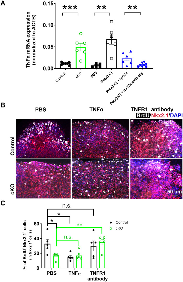
Fig. 6. Elevated TNFα mediates Gpr56 cKO–induced MGE proliferation deficits.
(A) TNFα mRNA was significantly elevated in E14.5 microglia isolated from Gpr56 cKO and MIA fetal brains. IL-17a–neutralizing antibodies significantly attenuated microglial TNFα mRNA elevation in MIA-treated fetal brains. n = 5 to 8. ACTB, actin beta. (B) Representative images of BrdU and Nkx2.1 in ex vivo culture slices. (C) The percentage of BrdU+ cells in the MGE were significantly decreased in Gpr56 cKO slices compared to control slices under PBS treatment. In control slices, TNFα treatment significantly decreased the percentage of BrdU+ progenitors in the MGE, while treatment with TNFR1-neutralizing antibodies had no effect (P > 0.99). In Gpr56 cKO slices, TNFR1-neutralizing antibody treatment significantly increased MGE proliferation, while TNFα treatment did not alter MGE proliferation (P > 0.99). Control: n = 5; cKO: n = 6. Unpaired t test for (A) and two-way ANOVA and Bonferroni’s post hoc test for (C). *P < 0.05, **P < 0.01, and ***P < 0.001. Data are presented as means ± SEM.

