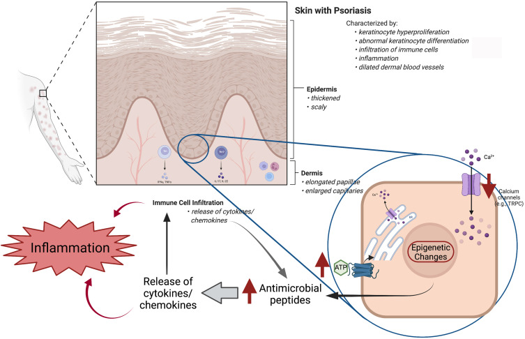
Figure 2.
The structure of psoriatic skin with intrinsic differences between psoriatic and normal keratinocytes highlighted. On top is shown a schematic of psoriatic skin illustrating the scaly stratum corneum and the overall thickened epidermis, including elongated papillae and rete ridges with dilated capillaries in the dermis, as well as the immune cell infiltration and inflammation. In the inset, a keratinocyte from the basal layer of the epidermis is enlarged to illustrate intrinsic differences observed in psoriatic versus normal keratinocytes. The changes in psoriatic keratinocytes are indicated by the red arrows and include: abnormal calcium metabolism via decreased capacitative calcium influx [for example, decreased transient receptor potential canonical (TRPC) channel levels] and an increased cytosolic calcium response to ATP, as well as enhanced secretion of anti-microbial peptides, apparently upregulated at least in part as a result of epigenetic changes (e.g., hypomethylation of the S100A9 gene). All of these alterations in psoriatic keratinocytes amplify the inflammatory immune feedback loop that initiates and sustains chronic inflammation in psoriasis. Created using Biorender.com.

