Figure 6.
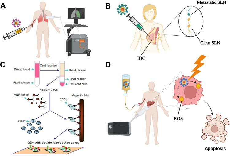
Some potential applications of QDs in clinics. (A) Diagnostic imaging. QDs modified with specific targeting ligands can be injected intravenously to accumulate into the target organ, allowing its visualization. (B) Sentinel lymph node (SLN) detection. QDs can substitute the currently-used blue dye or radionuclide methods for the intraoperative detection of SLNs. Following peritumoral injection, QDs diffuse to the affected SLN(s) allowing accurate and safe detection of SLNs metastases. (C) Detection of micrometastases. Blood samples from cancer patients are fractioned by gradient centrifugation to collect PBMC and CTCs, followed by their mixing with magnetic nanoparticles that are labeled with anti-pan-ck antibody to enrich lung cancer epithelial cells. The micrometastatic cells conjugated to the magnetic nanoparticles are separated using a magnetic field, incubated with double antibody-labelled QDs, and detected under a fluorescence microscope. The figure (6C) is reproduced from Wang Y, Zhang Y, Du Z, Wu M, Zhang G. Detection of micrometastases in lung cancer with magnetic nanoparticles and quantum dots. Int J Nanomedicine. 2012;7:2315–2324. With a permission from Dove Medical Press, copyright 2012.81 (D) Cancer photodynamic therapy. Following administration of QDs, they tend to accumulate into the tumor site via passive or active targeting. Local irradiation of the tumor site induces the generation of ROS that kill the tumor cells. Created by BioRender.com.
Abbreviations: IDC, invasive ductal carcinoma; SLNs, sentinel lymph nodes; CTCs, circulating tumor cells; MNPs, magnetic nanoparticles; Pan-ck, pan-cytokeratin; PBMC, peripheral blood mononuclear cell. ROS, reactive oxygen species.

