Figure 5.

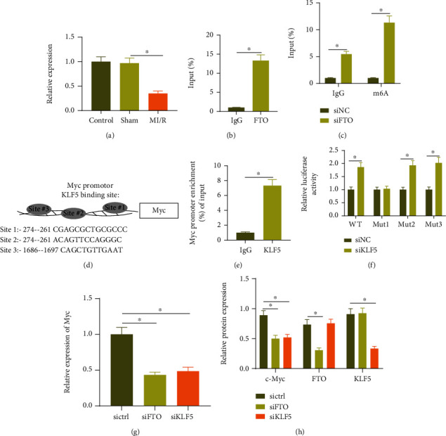
FTO and KLF5 promote Myc expression in cardiomyocytes: (a) RT-qPCR detection of Myc expression in the myocardial tissue of sham-operated mice and MI/R mice; (b) RIP analysis using FTO specific antibody in cardiomyocytes; (c) MeRIP adopted to analyze the m6A modification of Myc mRNA in the cardiomyocytes of each group; (d) bioinformatics analysis of the top three binding sites of KLF5 and Myc promoter; (e) ChIP detected the enrichment of KLF5 in the Myc promoter region in cardiomyocytes; (f) dual-luciferase assay detected the targeting relationship between KLF5 and Myc; (g) RT-PCR was employed to detect the expression of Myc mRNA in the cardiomyocytes of each group; (h) Western blot analysis was used to detect the protein of FTO, KLF5, and Myc in the cardiomyocytes of each group. Measurement data were expressed as the mean ± standard deviation. Data in compliance with normal distribution and homogeneity of variance between two groups were compared using an unpaired t-test. Comparisons among multiple groups were conducted by one-way ANOVA or repeated measurement ANOVA with Tukey's post hoc test. p < 0.05 was indicative of statistical significance. The cell experiment was repeated three times.
