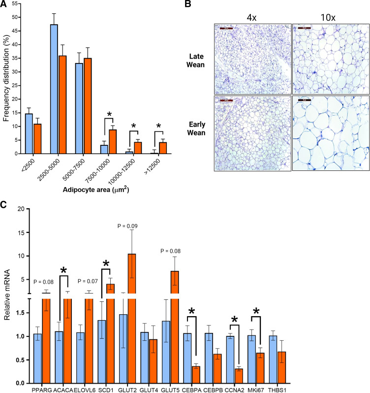
Figure 9.
Increased adipocyte size and decreased proliferation in visceral adipose tissue from early-weaned pigs. A: H&E staining of perirenal-abdominal adipose tissue from early- and late-weaned pigs at day 70 of age. Frequency distribution (percentage) of adipocyte area. B: representative histological images of perirenal adipose tissue at ×4 and ×10 magnifications. C: relative gene expression of adipose tissue. Values represent means ± SE for n = 6–8 pigs/weaning age group. *P < 0.05, LW vs. EW, Student’s t test. EW, early weaned; H&E, hematoxylin-eosin; LW, late weaned.

