Fig. 1.
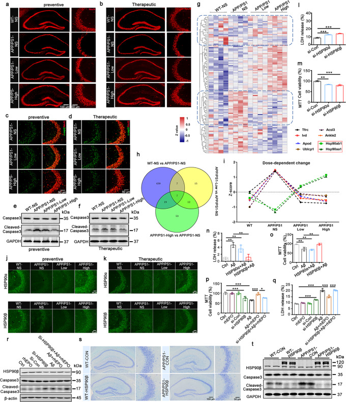
rhEPO attenuated neuronal loss via HSP90β. a, b Representative images of NeuN immunostaining in the hippocampus of the mice in the prevention (a) and treatment experiments (b). c, d Representative images of Neuronal apoptosis detected by immunofluorescence using anti-cleaved-caspase3 (c-cap3) antibody in hippocampal CA3 in the prevention (c) and treatment experiments (d). e, f Western blotting for Caspase3 and Cleaved-caspase3 in the hippocampal CA3 in the prevention (e) and treatment experiments (f). g The heat map of total differentially expressed (DE) proteins after rhEPO treatment, red represents high expression abundance, and dark blue represents low expression abundance. h Venny analysis to find the shared proteins among different compared groups after rhEPO treatment. i The dose-dependent-change of shared proteins among compared groups of rhEPO-treated mice vs untreated mice analyzed by Venny analysis. The proteins abundance was shown as Z-score. j, k Representative images of HSP90α and HSP90β immunostaining in the hippocampal CA3 in the prevention (j) and treatment experiments (k). l, m Knockdown of HSP90α or HSP90β in N2a cells promoted cell apoptosis detected by LDH assay (l) and MTT (m). n, o Overexpression of HSP90β in N2a cells attenuated cytotoxicity induced by Aβ detected by LDH assay (n) and MTT (o). p–r Knockdown of HSP90β attenuated rhEPO-induced cytoprotective effects against Aβ in N2a cells, which were detected by LDH assay (p), MTT (q), and cleaved-caspase3 was detected by Western blotting (r). s, t AAV-HSP90β-mcherry virus (1.5 × 10¹³v.g/ml) was stereotaxically injected into the hippocampal CA3 of 7.5-month-old C57 and APP/PS1 mice, neuronal number detected by Nissl staining in the hippocampal CA3 region (s), caspase3 or cleaved-caspase3 level was detected by Western blotting. Data were presented as mean ± SD. *p < 0.05, **p < 0.01, ***p < 0.001. N = 3 per group

