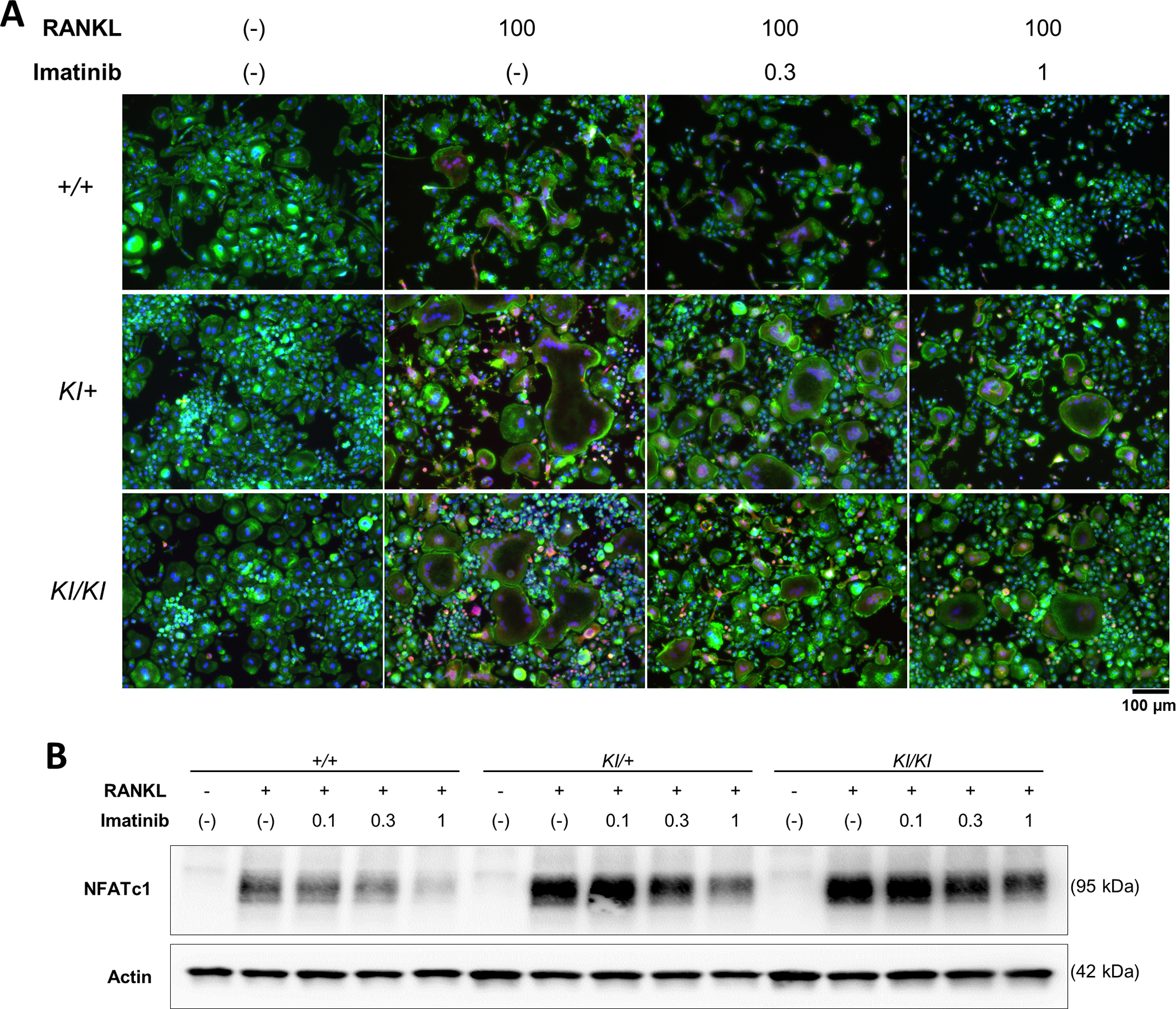
Figure 3. Imatinib decreases osteoclast formation via suppressed NFATc1 expression.

Bone marrow cells were isolated from Sh3bp2+/+, Sh3bp2KI/+, and Sh3bp2KI/KI mice. Non-adherent bone marrow cells were seeded at a density of 1.0 × 10⁵/mL. After a 2-day preculture with M-CSF (25 ng/mL), BMMs were stimulated with RANKL (50 ng/mL) in the presence or absence of imatinib for 72 h. (A) Fluorescent staining of RANKL-stimulated BMMs. Cells were fixed with 2% PFA/PBS. Actin and nuclei were visualised with Alexa Fluor-488-conjugated phalloidin and 4’, 6-diamidino-2-phenylindole, respectively. NFATc1 was stained with an anti-NFATc1 antibody, followed by Alexa Fluor-555-conjugated anti-mouse antibody. Actin, nuclei, and NFATc1 were indicated in green, blue, and red, respectively. Original magnification, 20×. (B) Immunoblot analysis of NFATc1. Protein samples were collected at 48 h after the RANKL treatment. Actin was used as a loading control. SH3BP2, SH3 domain-binding protein 2; +/+, wild-type; KI, knock-in; BMMs, bone marrow-derived macrophages; RANKL, receptor activator of nuclear factor-κB ligand; PFA/PBS, paraformaldehyde/phosphate-buffered saline.
