
Figure 5. Imatinib does not rescue the osteopenic phenotypes of Sh3bp2KI/KI mice.
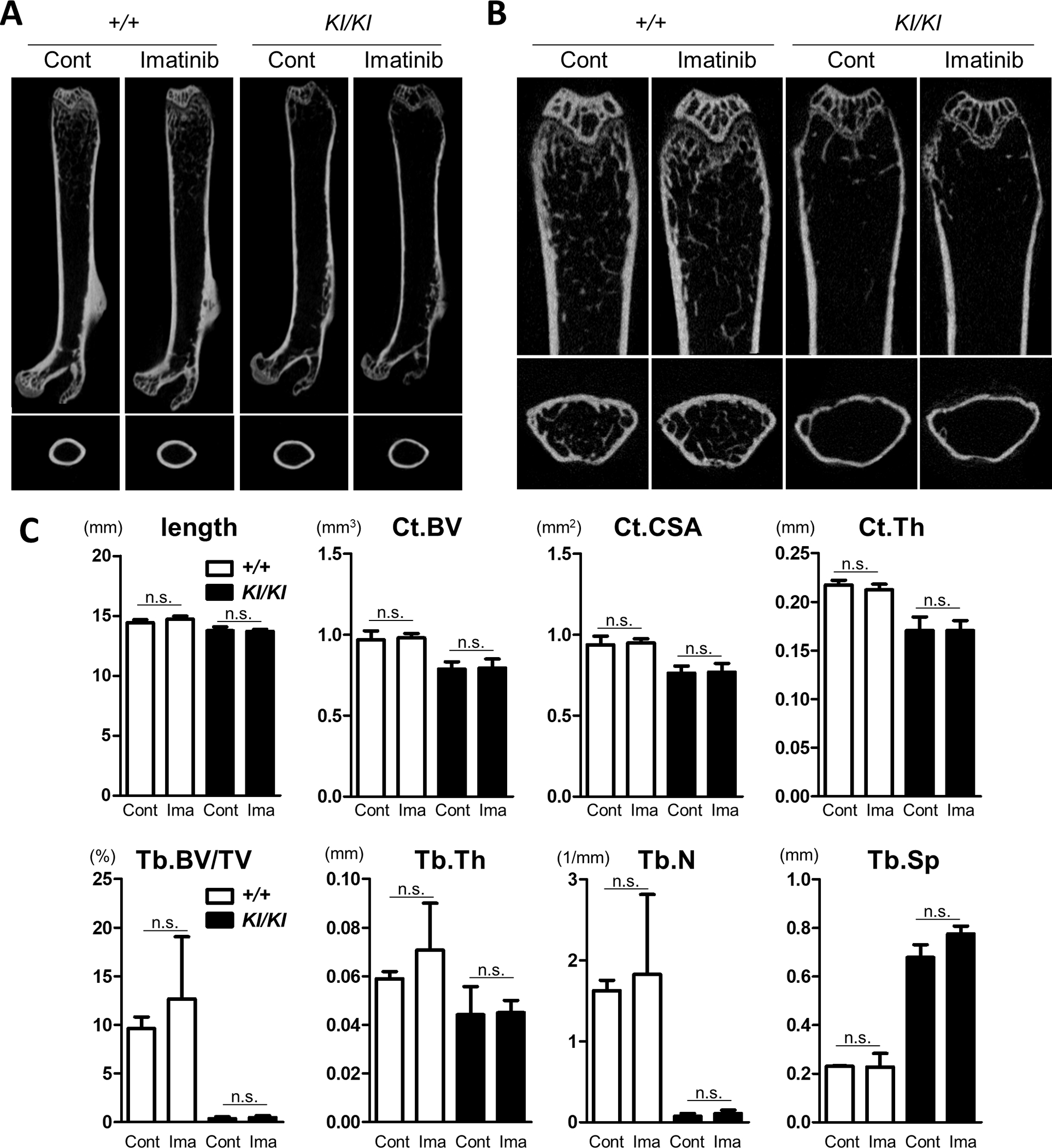
Imatinib was administered intraperitoneally to 10-week-old female Sh3bp2+/+ and Sh3bp2KI/KI mice daily 5 times per week for 4 weeks (n = 4–5/group). PBS was administered to the control mice. At the end of the experiments, the femurs were collected and subjected to micro-CT analysis. (A) Representative images of the whole femurs and cortical bones at the midshaft. (B) Representative images of the trabecular bone in the distal femurs. (C) Bone properties. Bone lengths were measured. Cortical bone volume (Ct.BV), cortical cross-sectional area (Ct.CSA), and cortical thickness (Ct.Th) of the midshaft of femurs were determined by micro-CT. Trabecular bone volume per total volume (Tb.BV/TV), trabecular thickness (Tb.Th), trabecular number (Tb.N), and trabecular separation (Tb.Sp) were determined at the distal side of the femurs. Values are presented as the mean ± SD. Note: * p < 0.05; n.s. = not significant. SH3BP2, SH3 domain-binding protein 2; +/+, wild-type; KI, knock-in; Cont, control; Ima, Imatinib; PBS, phosphate-buffered saline; CT, computed tomography.