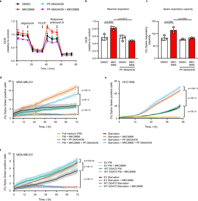
Fig. 5. MKC8866 sensitizes cells to starvation through regulation of DGAT2.
a–c MDA-MB-231 cells were treated with vehicle or 20 μM MKC8866 in the presence or absence of 2 µM PF-06424439 for 6 days (n = 3 biologically independent experiments). a–c Seahorse extracellular flux analysis of oxygen consumption rate (OCR) following the indicated injections normalized to vehicle. Data plotted to demonstrate the changes in mitochondrial b maximal respiration and c spare respiratory capacity. Indicated p values based on comparison of multiple groups using one-way ANOVA with Bonferroni’s multiple comparisons post hoc tests. Values with p < 0.05 are considered statistically significant. Data are presented as mean values ± s.d. d MDA-MB-231 cells (n = 4 biologically independent experiments), e HCC1806 (n = 4 biologically independent experiments) and f MDA-MB-231 cells stably expressing EV or WT DGAT2 (n = 3 biologically independent experiments) were treated with vehicle or 20 μM MKC8866 in the presence or absence of 2 μM PF-06424439. After 6 days, culture medium was replaced with a complete medium or Hanks’ balanced salt solution. Kinetics of cell death for up to 72 h was expressed as the percentage of cells that were Sytox Green positive. Indicated p values based on two-way ANOVA with Bonferroni’s multiple comparisons post hoc tests. Values with p < 0.05 are considered statistically significant. Data are presented as mean values ± s.e.m. Source data are provided as a Source Data file.

