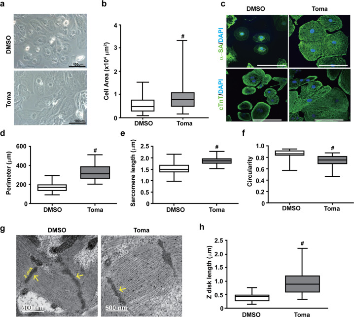
Fig. 2. Effects of tomatidine treatment on the morphology of human embryonic stem cell-derived cardiomyocytes.
a, b Effects of tomatidine treatment on the morphology of hESC-CMs. a Bright field images of the control CMs and Toma-CMs on Day 30. Scale bar = 100 μm. b Quantification of the cell area of the control CMs and Toma-CMs. c–f Immunocytochemistry images (c) and quantification of the measurements of the perimeter (d), sarcomere length (e), and circularity (f) of the control CMs and Toma-CMs (n = 77). Representative TEM images (g) and quantification of the measurement of the z disk length (h) in the control CMs and Toma-CMs. Data are shown as the mean ± S.D. (n = 33). *p < 0.05; ‡p < 0.01; #p < 0.001.

