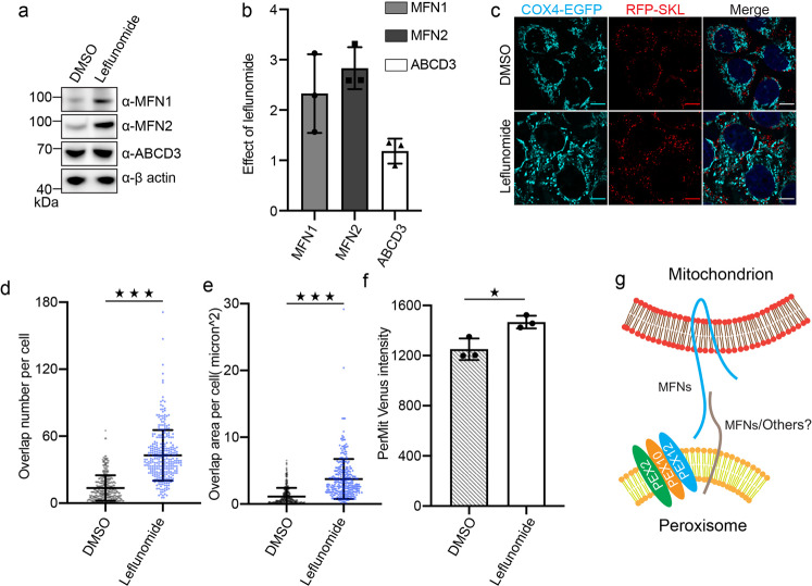
Fig. 7. Upregulation of endogenous mitofusins stimulates mitochondrion-peroxisome contacting.
a Immuno-blots of MFN1, MFN2, and ABCD3 in cells treated with/without leflunomide. b Fold change of protein levels on immune-blots (a), quantified with ImageJ (n = 3, error bars represent standard deviation). c Representative fluorescence images of the HeLa cells stably expressing COX4-EGFP and RFP-SKL, treated with either DMSO or leflunomide (50 μM, 48 h). d, e Quantification of peroxisome and mitochondrion overlap in images collected in (c). Scatter dot plots showing peroxisome and mitochondria overlap number and area in HeLa cells incubated with DMSO (n = 348 cells) and 50 μM leflunomide (n = 310 cells) respectively for 48 h. ***p < 0.001. Mean with SD. P values calculated via unpaired Student’s t-test (two-tailed). f The Venus fluorescence intensity in cells treated with either DMSO or 50 μM leflunomide were analyzed with flow cytometry. At each condition, the mean fluorescence intensity (MFI) is calculated from 20,000 to 60,000 cells. The experiment was repeated for three times. The columns represent average MFI (mean fluorescence intensity) with SD. *p < 0.05, calculated via unpaired Student’s t-test (two-tailed). g A working model for MFN mediated PerMit contacting.

