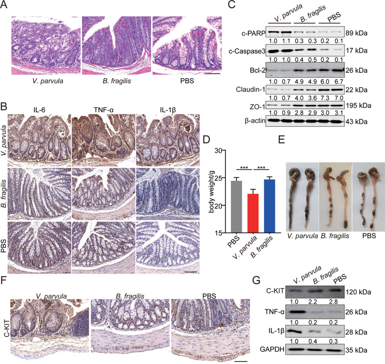
Fig. 1. The promoting effects of V. parvula overabundance on intestinal inflammation in vivo.
A Representative photomicrographs showing worse histologic injury, more crypt abscesses and more distribution of immune cells in the distal colon of mice treated with V. parvula. Scale bars: 100 μm. B Higher levels of inflammatory cytokines (IL-6, IL-1β and TNF-α) in distal colon of mice treated with V. parvula. Scale bars: 100 μm. C Western blotting assays showing more apoptosis and impaired tight junction induced by V. parvula than B. fragilis or PBS in distal colon tissues. D Body weight of mice treated with V. parvula is lighter compared to B. fragilis or PBS. E Representative images showing more severe intestinal dysmotility and colonic dilatation in mice of V. parvula group compared to mice of B. fragilis or PBS group. F Representative photomicrographs of IHC staining showing less C-KIT+ ICC in mice of V. parvula group compared to mice of B. fragilis or PBS group. Scale bars: 100 μm. G Western blotting assays showing more IL-1β and TNF-α and less C-KIT induced by V. parvula than B. fragilis or PBS in distal colon tissues. All data are expressed as mean ± SD. ***P < 0.001.

