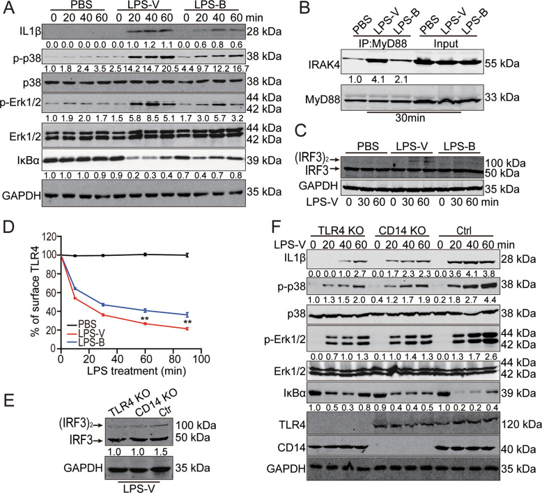
Fig. 5. LPS-V activated macrophages through TLR4 signaling.
A Western blotting assays showing IL-1β and TNF-α as well as MyD88-dependent changes in IκBα levels, p-p38, and p-ERK1/2 at indicated time points of treatment with LPS (1 μg/ml) in BMDMs. B IP and Western blot analysis of the complex of MyD88 and IRAK4 30 min post treatment with LPS (1 μg/ml) in BMDMs. C Western blotting assays using Native-PAGE electrophoresis indicating active (dimerized) IRF3 at indicated time points of treatment with LPS (1 μg/ml) in BMDMs. D Mean fluorescence intensity (MFI) of TLR4 receptor staining at each time point of treatment with LPS (1 μg/ml) in BMDMs. Data are expressed as mean ± SD. **P < 0.01. E TLR4 or CD14 KO decreased active (dimerized) IRF3 in macrophages differed from THP1 treated with LPS-V. F Western blotting assays showing that TLR4 or CD14 KO decreased IL-1β and TNF-α as well as MyD88-dependent IκBα levels, p-p38, and p-ERK1/2 at indicated time points of treatment with LPS (1 μg/ml) in macrophages differed from THP1.

