Figure 5.

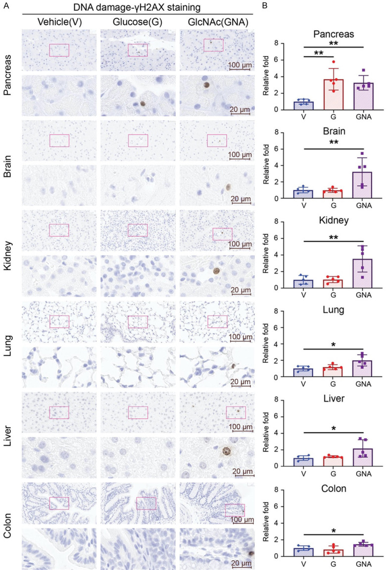
High dose of GlcNAc treatment induces DNA damage in tissues. Five male mice of each group were treated with glucose (G, 15000 mpk) or GlcNAc (GNA, 7500 mpk) or vehicle control (water) for 7 days. Glucose and N-Acetylglucosamine were dissolved in water and administered to mice three times daily by oral gavage at 10 μl/g. (A) Representative images and (B) quantifications of γ-H2AX staining by IHC of pancreas, brain, kidney, lung, liver, and colon from each group. (A) Upper panel: 40× magnification. Scale bar, 100 μm. Lower panel: 200× magnification. Scale bar, 20 μm. (B) Quantitative bar graphs of the relative folds of the percentage of γH2AX positive cells. Each dot represents the datum of one mouse. Data are normalized to that from mice treated with vehicle control. Values show mean ± SEM. *P<0.05; **P<0.01 (two-tailed Student’s t-test).
