
FIGURE 3.
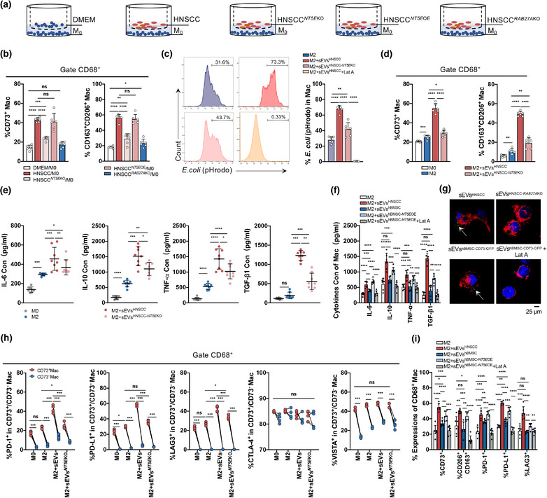
The effect of CD73 in sEVs derived from HNSCC cells on the function of macrophages. (a) Macrophages were cocultured with DMEM or two HNSCC lines (SCC25, HN6) with or without NT5E/RAB27AKO for 24 h. HNSCC NT5E OE, referred to overexpression of NT5E performed on HNSCC NT5E KO cells. (b) Flow cytometry analysis for percentage of CD73+ macrophages and M2 macrophages (CD163+CD206+) in coculture system. (c–i) The sEVs (50 μg) were cocultured with macrophages (1 × 10⁶) for 24 h. The sEVsHNSCC derived from two HNSCC lines: SCC25 and HN6. The sEVshBMSC‐ NT5E OE derived from hBMSC cells with NT5E overexpression. Latrunculin A (Lat A, 30 μM) was used as the inhibitor of sEVs uptaken. (c) Compromised phagocytosis of M2 macrophages treated with or without sEVs or Lat A. The percentage of pHrodo dyes of M2 macrophages was analyzed by flow cytometry. (d) The percentage of CD73+ macrophages and M2 macrophages after macrophages cocultured with sEVs. (e, f) Concentration of IL‐6, IL‐10, TNF‐α, and TGF‐β1 levels in macrophages conditional medium after sEVs education by ELISA. (e)The sEVs were derived from HNSCC cells or HNSCC NT5E KO cells. (f) The sEVs were derived from HNSCC, hBMSC or hBMSC NT5E OE cells with or without Lat A (30 μM). (g) Macrophages were cocultured with anti‐CD73‐FITC labelled sEVs from HNSCC cell lines control or RAB27AKO, CD73‐GFP labelled sEVs from hBMSC treated with or without Lat A were cocultured with macrophages for 1 h, and Laser Scanning Confocal Microscopy was used to analyze the internalization of HNSCC‐derived sEVs into macrophages (Scale bar = 25 μm). (h) Flow cytometry analysis for differential expression of immune check point (PD‐1, PD‐L1, LAG3, CTLA‐4, VISTA) comparing M0 and M2 macrophages which were educated with sEVs from HNSCC cells (h) or hBMSC with or without Lat A (i). Data were analysed by Mann‐Whitney test (ns, no significant difference, *p < 0.05, **p < 0.01, ***p < 0.001, ****p < 0.0001)