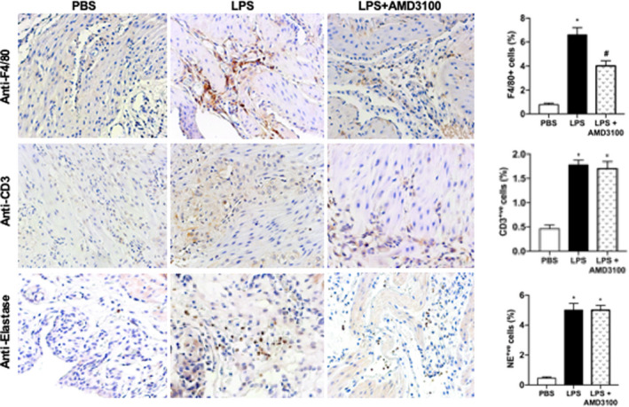
FIGURE 4.

LPS‐stimulated engraftment of immune cells into the uterus while AMD3100 inhibited macrophage engraftment. Representative IHC images showing that the number of immune cells engrafted were increased by LPS treatment compared to PBS treated controls. Macrophages (anti‐F4/80); T cells (anti‐CD3) and leukocytes (anti‐elastase) are all increased by LPS treatment, however, only macrophages are decreased after AMD3100 treatment. On the right quantitative analyses demonstrates the significant increase in F4/80+ve, CD3+ve and elastase+ve cells in LPS‐treated mice. AMD3100 inhibited the macrophage (F4/80+ve) engraftment induced by LPS. Results shown as mean ± SE. *p < 0.05 versus PBS while # p < 0.05 versus LPS. Original magnification 400×. IHC, immunohistochemistry; LPS, lipopolysaccharide; PBS, phosphate buffered saline; SE, standard error
