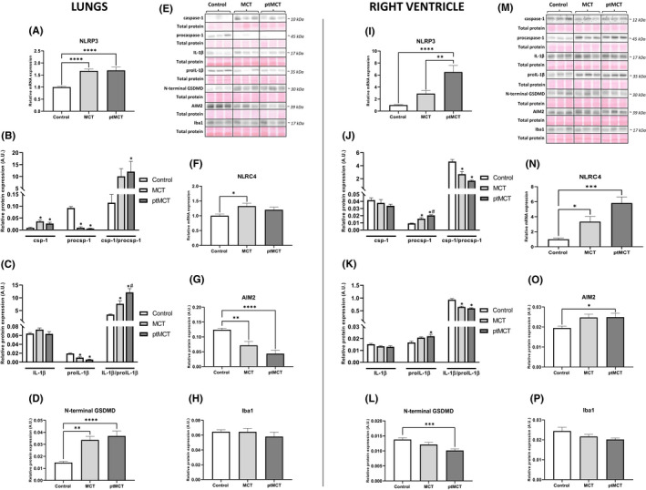
FIGURE 5.

Analysis of inflammasomes and pyroptotic signaling in the lung (left panel) and RV (right panel) tissue. Relative mRNA expression of NLRP3 (A, I) and NLRC4 (F, N), relative protein expression (B‐D, G, H, J–L, O, P) and representative immunoblots and total protein staining (E, M) of csp‐1, procsp‐1 (B, J), IL‐1β, pro‐IL‐1β (C, K), N‐terminal GSDMD (D, L), AIM2 (G, O) and Iba1 (H, P) in control group, monocrotaline group (MCT) and prematurely terminated monocrotaline group (ptMCT). Data are presented as mean ± SEM; n = 8–10 per group; *p ˂ 0.05 vs. Control; # p ˂ 0.05 vs. MCT
