. 2018 Jan 25;8(9):4624–4633. doi: 10.1039/c7ra13454a

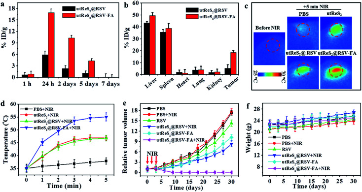
Fig. 7. (a) Biodistribution of the nanoparticles in the tumor tissue post-injection with utReS2@RSV and utReS2@RSV–FA. (b) Biodistribution of the nanoparticles in the major organs (i.e., heart, liver, spleen, lung, kidney) as well as tumors at 24 h post-injection with utReS2@RSV and utReS2@RSV–FA. (c) The thermal images of tumor-bearing mice at 24 h post-injection of PBS, utReS2, utReS2@RSV and utReS2@RSV–FA under 5 min NIR irradiation (808 nm, 1 W cm−2), respectively. (d) The temperature statistical results of tumor regions of tumor-bearing mice post tail vein injection of PBS, utReS2, utReS2@RSV and utReS2@RSV–FA at 24 h under 5 min NIR irradiation (808 nm, 1 W cm−2), respectively. (e) The growth profile of HepG2 tumors after intravenous injection of PBS, RSV, utReS2@RSV and utReS2@RSV–FA with or without three cycles of NIR irradiation (5 min, 808 nm, 1 W cm−2) once a day. (f) Body weight of tumor-bearing mice during 30 days of treatment.

Fig. 7