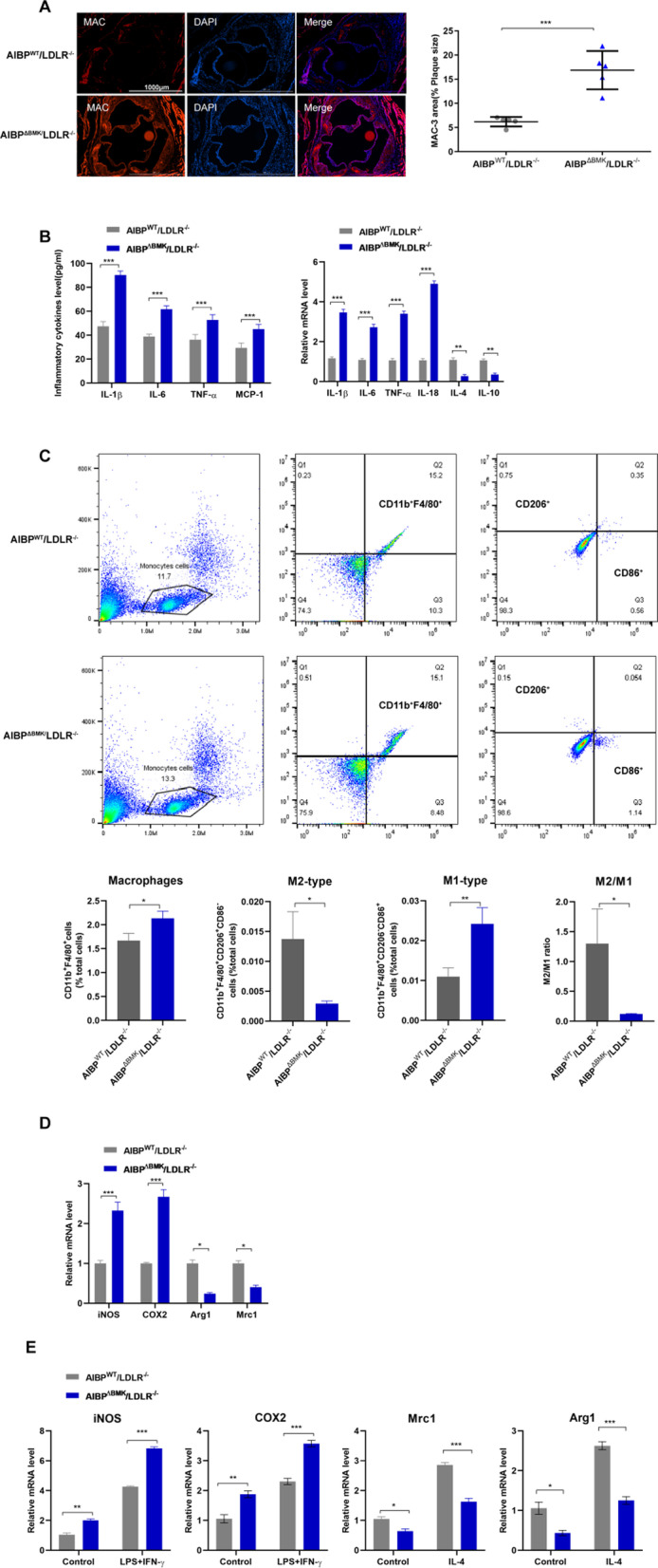
Fig. 2.

AIBPΔBMK promotes macrophage infiltration and proinflammatory polarization in LDLR−/− mice. A Representative images of Mac3 staining in aortic roots isolated from AIBPΔBMK/LDLR−/− and AIBPWT/LDLR−/− mice fed a high-fat diet (n = 3). Scale bar = 100 μm. B ELISA and qPCR were used to detect the protein and mRNA levels of inflammatory cytokines in the aorta (n = 5). C Flow cytometry analysis of macrophage infiltration and numbers of M1/M2-type macrophages in the blood of AIBPΔBMK/LDLR−/− and AIBPWT/LDLR−/− mice after 8 weeks of HFD consumption. n = 4–5 mice per group. D qPCR analysis of the mRNA levels of macrophage phenotype markers in aortas from AIBPΔBMK/LDLR−/− and AIBPWT/LDLR−/− mice (n = 5). E qPCR analysis of the mRNA levels of M1 and M2 markers in MPMs from AIBPΔBMK/LDLR−/− and AIBPWT/LDLR−/− mice after 8 weeks of HFD consumption that were stimulated with LPS + IFN-γ or IL-4 (n = 5). Data are presented as the means ± SD. *P < 0.05, **P < 0.01, and ***P < 0.001; one-way ANOVA followed by the Newman–Keuls test
