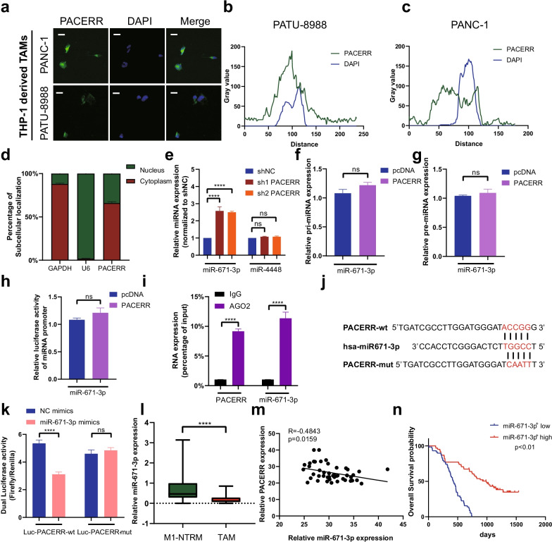
Fig. 4.
LncRNA-PACERR functions as a ceRNA to sponge miR-671-3p in TAMs. A Fluorescence in situ hybridization (FISH) of LncRNA-PACERR (green) in THP-1-derived TAMs (co-cultured with PATU-8988 or PANC-1). DAPI staining (blue) shows the nuclei. Scar bar: 10 μm. B, C Grey value of LncRNA-PACERR and DAPI on the FISH from THP-1 derived TAMs. Green represents LncRNA-PACERR. Blue represents DAPI. D Expression levels of LncRNA-PACERR in the cytoplasm and nucleus in THP-1-derived TAMs. E Expression of two potential target miRNAs (miR-671-3p and miR-4448) in THP-1 derived TAMs after LncRNA-PACERR knocked down. F, G Expression of pri-miR-671-3p (F) and pre-miR-671-3p (G) in THP-1 derived TAMs transfected with empty control or pcDNA-LncRNA-PACERR. H Promoter luciferase activity of miR-671-3p in 293-T cells overexpressing LncRNA-PACERR. I RIP assay was performed using rabbit AGO2 and IgG antibodies in THP-1 derived TAMs. Relative expression levels of LncRNA-PACERR and miR-671-3p were determined by qRT-PCR. J, K Dual luciferase activity in 293-T cells co-transfected with LncRNA-PACERR wild-type or mutant sequence and miR-671-3p mimics. L Expression of miR-671-3p in 46 pairs of TAMs and M1-NTRMs from PDAC patients. M Correlation analysis between LncRNA-PACERR and miR-671-3p using expression data from 46 pairs of TAMs. N Kaplan–Meier survival curve presenting the overall survival of 110 PDAC patients, grouped according to the extent of miR-671-3p+ TAMs infiltration

