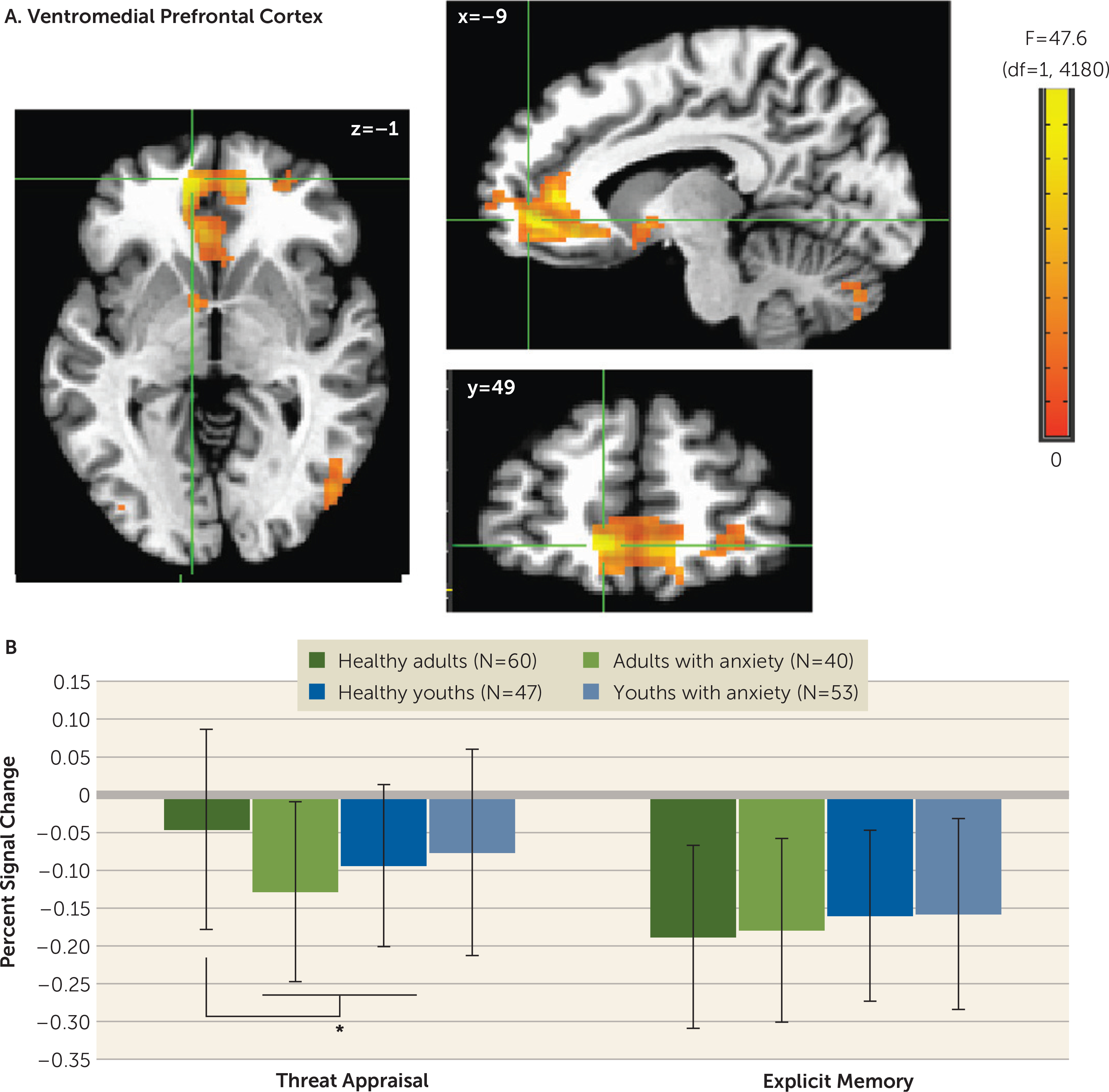
FIGURE 2. Task-related activation in the ventromedial prefrontal cortex among youths and adults with anxiety disorders and healthy subjectsa.

a Whole-brain analyses of task-related activation revealed a significant interaction of anxiety diagnosis, age, and attention condition in the ventromedial prefrontal cortex (vmPFC) (panelA). Images are shown in neurological convention (i.e., left is left) and thresholded at F>10.76, df=1, 4180, p<0.001, cluster size >57 voxels (890.625 mm3). To decompose the complex interaction effects, mean extracted values (panel B) for this cluster are plotted separately by attention condition (threat appraisal, explicit memory) and group, based on anxiety diagnosis (healthy, anxiety) and age (median split: adults, youths). In the graph, the y-axis shows extracted vmPFC percent signal change averaged across participants in each group. Error bars indicate standard deviation.
*p<0.05.
