. 2018 Feb 9;8(12):6460–6470. doi: 10.1039/c7ra13062g

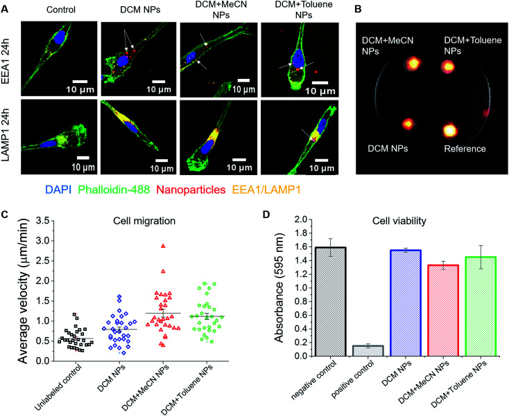
Fig. 5. Effect of nanoparticles on monocyte-derived DCs. (A) Confocal microscope images of nanoparticle uptake by DCs. Fluorescent signal coming from the nanoparticles partially overlaps with the signal of the early endosomal marker EEA1. Higher colocalization of nanoparticles was observed with the signal of late endosomal/lysosomal marker LAMP1. (B) 1H/19F MRI image showing the signal of 5 × 10⁶ cells loaded with different nanoparticles. Here, PFCE–PLGA particles, at 10 mg ml−1 concentration, were used as a reference. (C) Nanoparticles do not affect the migration of dendritic cells. The graph represents average velocity of unloaded cells and cells loaded with nanoparticles. (D) The graph shows the viability of cells after incubation with various nanoparticles for 72 hours. No significant influence on cell viability was observed. Error bars represent the standard deviation (n = 3).

Fig. 5