. 2018 Mar 1;8(17):9112–9119. doi: 10.1039/c7ra13777j

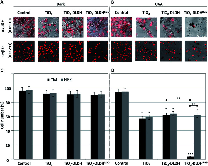
Fig. 4. Cytotoxic effect of the nanobiocomplex (A-NPs–DLDHRGD) in the presence or absence of UVA illumination. B16F10 or HEK293 cells were treated with TiO2, TiO2–DLDH or TiO2–DLDHRGD and assessed after an overnight by confocal microscopy (Leica SP5) under (A) dark conditions or (B) UVA illumination (1 h, 365 nm). Cells treated in medium lacking TiO2 or the DLDH forms, served as controls. The cell nuclei were stained red (Draq5). Experiments were repeated twice in triplicates. Next the same experiment was analyzed for cell counts under (C) dark conditions or (D) UVA illumination. Experiments were repeated 3 times in triplicates. Significance (*p < 0.05, **p < 0.005, ***p < 0.001) from control cells after uva illumination. Significance between the experiments groups (±RGD, ±αvβ3) are indicated by an horizontal line.

Fig. 4