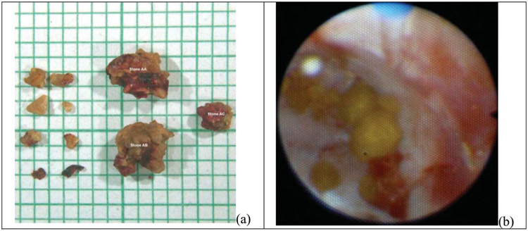
Figure 3.
Photograph of a 20% fragmented stone (a) and a 100% comminuted stone (b). In part a of figure, the largest 11 of 17 observed fragments were extracted and are shown on mm scale graph paper. The 2 largest fragments are approximately 4 mm in size. The stone was projected to fragment completely within 50 minutes. In part b of figure, all fragments were <2 mm, and mild clotting as well as bruising on the papilla is shown. This was scored as grade 1 injury.

