. 2018 Mar 1;8(17):9218–9222. doi: 10.1039/c8ra00774h

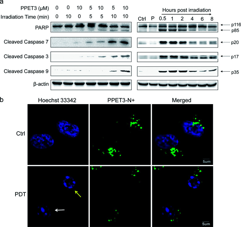
Fig. 4. (a) Western blot analysis of the expression of apoptosis-related proteins in HeLa cells. Left: HeLa cells were incubated with PPET3 (5 and 10 μM) and irradiated by white light for 5 or 10 min. The expression of the proteins was evaluated at 8 h post-treatment. Right: HeLa cells were incubated with PPET3 (5 μM) and irradiated by white light for 10 min. The expression of the proteins was evaluated at 0.5, 1, 2, 4, 6, and 8 h post-treatment, respectively. Ctrl: untreated cells, P: cells incubated with 5 μM PPET3 alone. (b) Confocal fluorescence images of PPET3 (5 μM)-treated HeLa cells after white light irradiation for 10 min. Cells were stained with Hoechst 33342. Scale bar: 5 μm.

Fig. 4