. 2018 Mar 12;8(18):10072–10080. doi: 10.1039/c7ra12503h

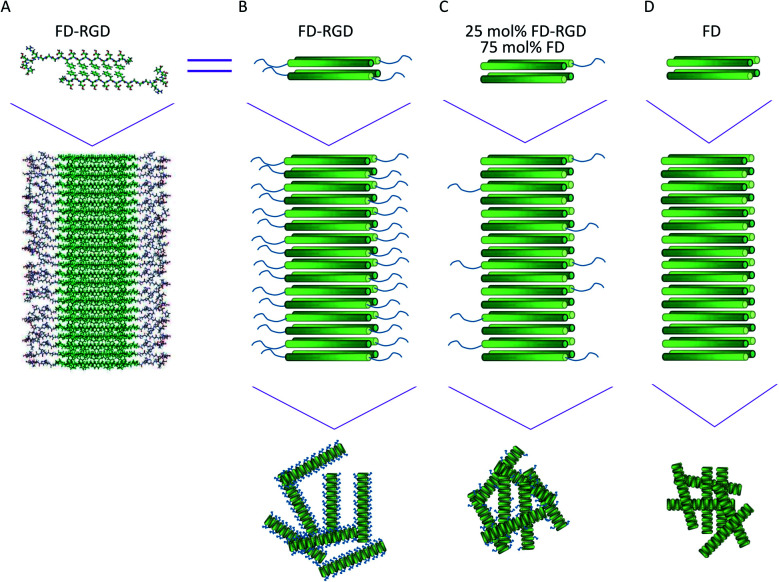
Fig. 1. Visualization of FD and FD-RGD assemblies. (A) Molecular models in rod presentation generated in Cerius2 (Accelrys Inc.); two FD-RGD peptides (top) composing the repeat motif of FD-RGD fibril (bottom). Both peptides are drawn with the FD and RGD sections in β-strand and random conformations. The β-strand conformation positions the hydrophobic, Phe side chains from both layers facing each other, while the hydrophilic side chains point to the surrounding aqueous phase. (B–D) The top scheme shows four peptides arranged in a bilayer that constitutes the fibril shown in the bottom scheme. The FD-β-strand section is drawn as a cylinder with a gradient color from the N- to C-termini (dark to bright, respectively) and the RGD-unordered part is depicted as a tail extending from the cylinder C-termini; (B) FD-RGD, (C) 25 mol% FD-RGD and (D) FD.

Fig. 1