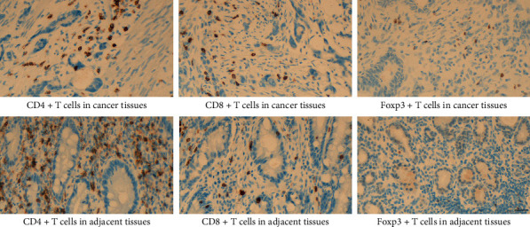
Figure 2.

Infiltration of CD4+T, CD8+T, and Foxp3+T cells in malignant tissues and normal adjacent tissues. CD4+T plus CD8+T cells were stained in yellow or brownish yellow on the cell membrane or cytoplasm, and the nuclear was brown or tan as Foxp3+T cells (×400), scale bar: 20 μm.
