. 2018 Mar 16;8(19):10620–10626. doi: 10.1039/c8ra00401c

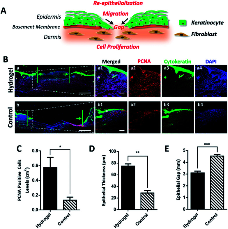
Fig. 5. Copolypeptide hydrogel promotes granulation tissue formation, keratinocyte cell proliferation and re-epithelialization. (A) Schematic diagram of the re-epithelialization process of wound healing, which is associated with cell proliferation and migration. Keratinocytes at the wound edge migrate towards the wound center to close the wound gap. (B) Immunofluorescent staining of cytokeratin (green) and proliferating cell nuclear antigen (PCNA, red). (a1)–(a4) and (b1)–(b4) are the minified images of selected areas in (a) and (b) with merged and split fluorescence channels. The scale bar in (a) and (b) is 1 mm, and 100 μm in magnified images. (C) Quantified PCNA positive cells levels on day 7 of the two groups. (D) Quantified epithelial thickness on day 7 of the two groups. (E) Quantified epithelial gap on day 7 of the two groups. Statistical differences were performed using one-way. analysis of variance (ANOVA). ***P value < 0.001, **P < 0.01, *P < 0.05, compared to the control group, n > 3.

Fig. 5