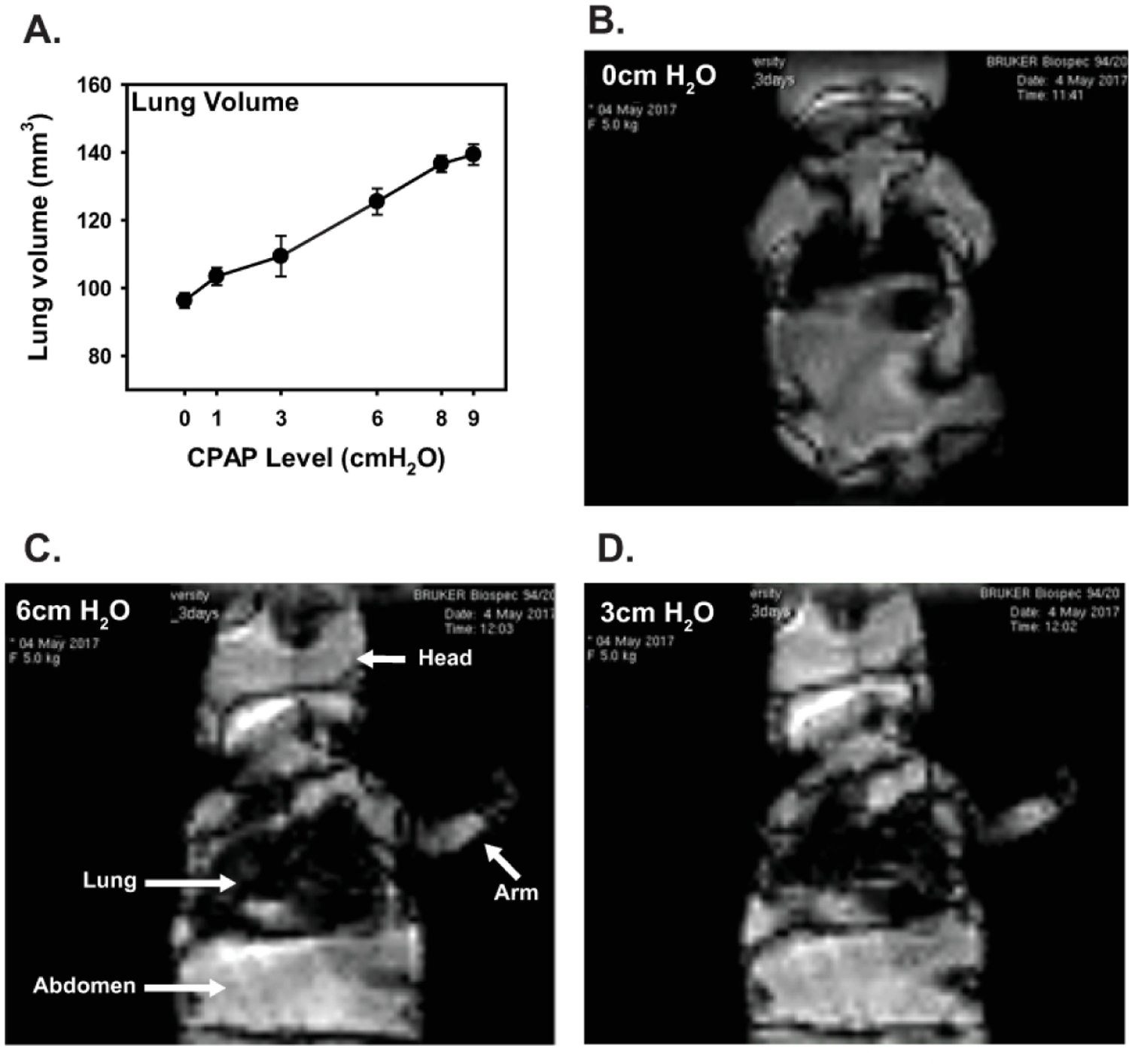
Figure 1:

Mean lung volume (A) and representative coronal FISP MRI images of the chest cavity from a 3 day old, unanethetized spontaneously breathing mouse while on various levels of acute CPAP (B-D, 0, 3, and 6 cmH2O, respectively). Values in A are means ± 1 SEM. Note, for imaging purposes, lung volumes were assessed at inflation pressures up to 9 cmH2O. For each scan, the animal was held at a given level of CPAP for ~60 seconds for image acquisition before CPAP was then increased to the next inflation pressure (n=4 mice).
