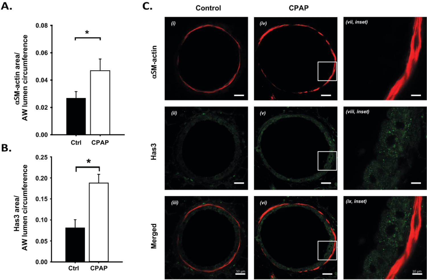
Figure 4:

Changes in epithelial HAS3 (A) and αSM-actin (B) immunoreactivity in P21 day old male mice, two weeks after CPAP (6 cmH2O) treatment ended. Note the increase in HAS3 (A) and αSM-actin (B) immunoreactivity following CPAP. Representative images of lung sections from a control (C, i-iii) and CPAP (iv-vi) treated mouse are provided, including high resolution images (inset, vii-ix) of the white box regions from the CPAP mouse. Note, HAS3 (green) was not co-localized with airway αSM-actin (red). *significantly different from control mice (p<0.05). (n=8 animals/group).
