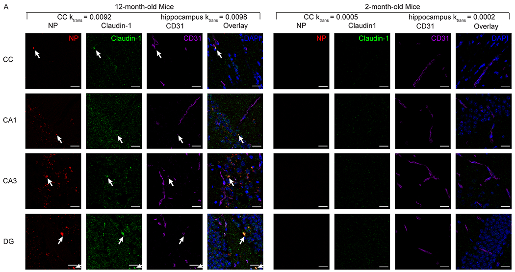
Figure 5.

Representative images of in vivo C1C2-NP (red) accumulation in the corpus callosum and hippocampus colocalize with the immunofluorescence staining for claudin-1 (green) and CD31 (purple). Blue DAPI staining indicates nuclei. Scale bar is 20 μm. NP accumulation and high claudin-1 co-localization with CD31 can be observed in the 12-month-old mice, but not in the 2-month-old mice.
