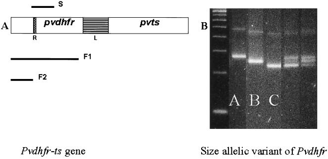
FIG. 1.
(A) Schematic representation of the pvdhfr-ts gene, with the linker region (L) and the repeat region (R) indicated. Three fragments were obtained by nested-PCR amplifications (S, F1, and F2) for further analysis. (B) Three S fragments of different sizes were observed and designated A, B, and C. In some samples, mixed infections were observed. Electrophoresis was performed in MetaPhor agarose in Tris-borate-EDTA, and the product was visualized by UV transillumination following ethidium bromide staining.

