. 2018 Apr 24;8(27):15229–15240. doi: 10.1039/c8ra02090f

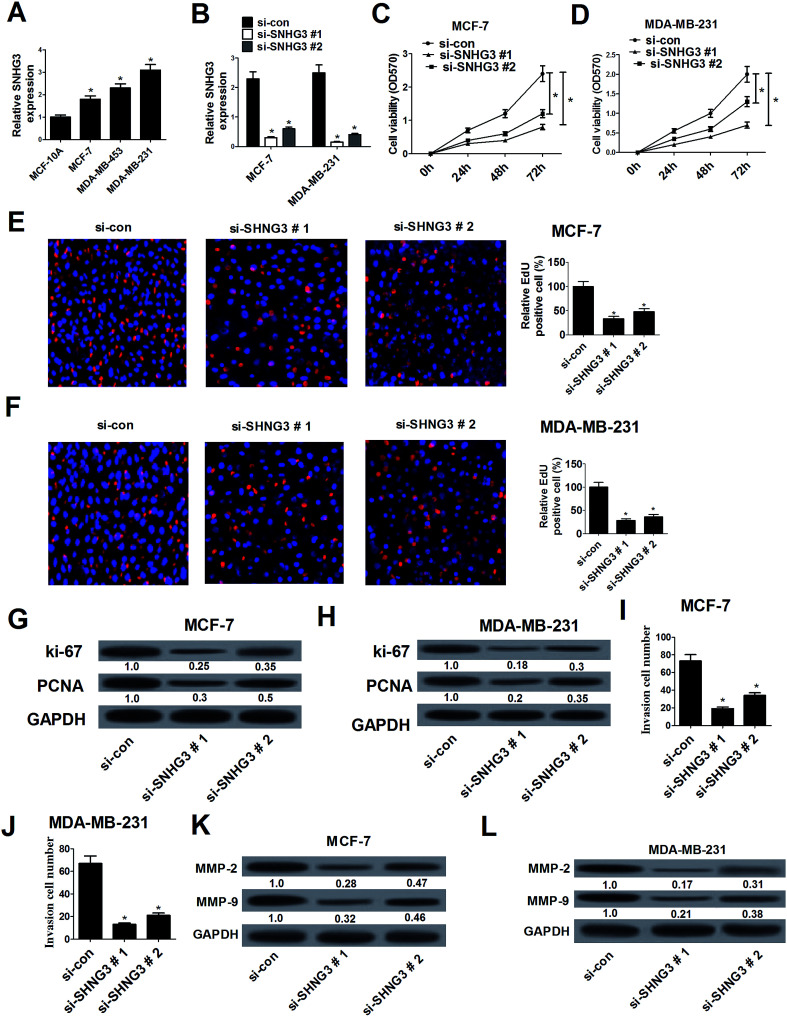
Fig. 2. SNHG3 silencing suppressed cell proliferation and invasion in breast cancer cells. (A) The expression level of SNHG3 in breast cancer cells (MCF-7, MDA-MB-453 and MDA-MB-231) and the normal breast epithelial cell line MCF-10A was estimated using qRT-PCR. (B) The expression level of SNHG3 in MCF-7 and MDA-MB-231 cells transfected with si-SNHG3#1, si-SNHG3#2 or si-con was evaluated using qRT-PCR. The MTT assay (C and D) and EdU proliferation assay (E and F) were performed to detect cell proliferation at 24 h, 48 h, and 72 h in MCF-7 and MDA-MB-231 cells transfected with si-SNHG3#1, si-SNHG3#2 or si-con. Western blot analysis was conducted to evaluate the protein levels of Ki-67 and PCNA in MCF-7 (G) and MDA-MB-231 (H) cells transfected with si-SNHG3#1, si-SNHG3#2 or si-con. Cell invasion ability was examined using the cell invasion assay in MCF-7 (I) and MDA-MB-231 (J) cells transfected with si-SNHG3#1, si-SNHG3#2 or si-con. The protein levels of MMP-2 and MMP-9 in MCF-7 (K) and MDA-MB-231 (L) cells transfected with si-SNHG3#1, si-SNHG3#2 or si-con were determined using western blot analysis. *P < 0.05.

Fig. 2