. 2018 Apr 24;8(27):15229–15240. doi: 10.1039/c8ra02090f

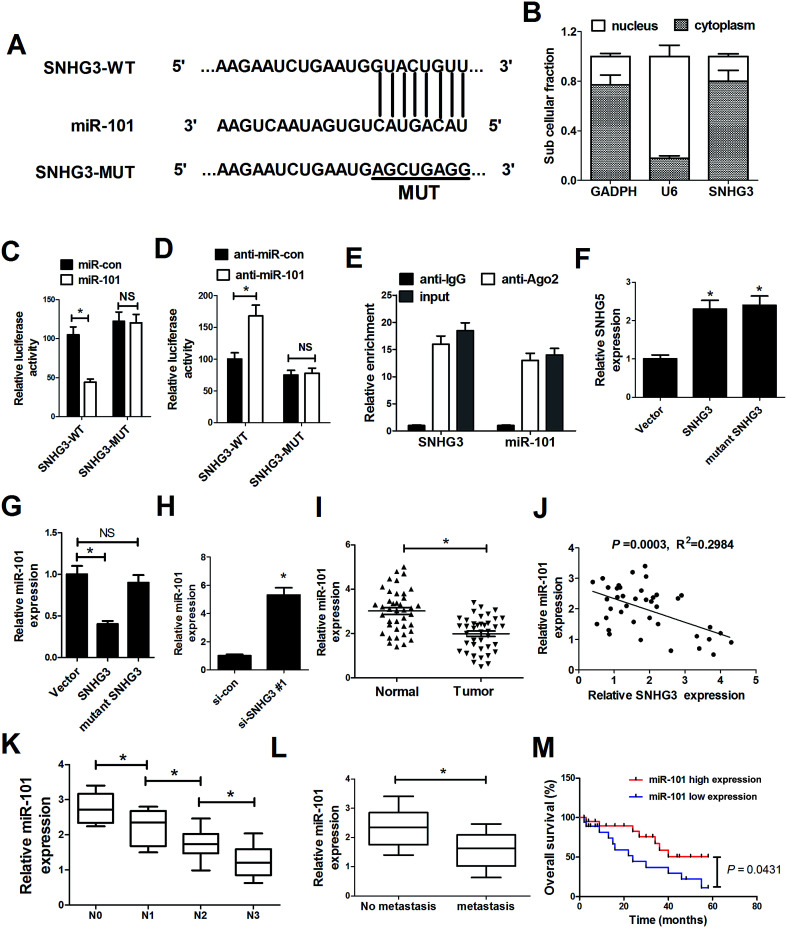
Fig. 3. SNHG3 functioned as a molecular sponge for miR-101 in breast cancer cells. (A) The luciferase reporter plasmids containing the predicted wild-type or mutated miR-101 in SNHG3. (B) Subcellular fractionation for SNHG3 in MCF-7 cells, where GAPDH and U6 were used as the controls. (C and D) A luciferase reporter assay was conducted to measure the luciferase activity in MCF-7 cells cotransfected with SNHG3-WT or SNHG3-MUT and miR-101, anti-miR-101, or matched controls. (E) Association between SNHG3 and miR-101 with Ago2 antibody. An anti-Ago2 RIP assay was performed in MCF-7 cell extract and the expressions of SNHG3 and miR-101 were detected using qRT-PCR. qRT-PCR was performed to detect the expressions of SNHG3 (F) and miR-101 (G and H) in MCF-7 cells transfected with SNHG3, mutant SNHG3, Vector, si-SNHG3#1, or si-con. (I) miR-101 expression in 42 paired breast cancer tissues and matched normal tissues. (J) The correlation between SNHG3 and miR-101 expression in breast cancer tissues. (K) The expression of miR-101 in different advanced pathological stages N0, N1, N2 and N3. (L) The expression of miR-101 in 31 breast cancer patients with distant metastasis and 11 breast cancer patients with no distant metastasis. (M) Kaplan–Meier curves and a log-rank test of overall survival for all breast cancer patients. *P < 0.05.

Fig. 3