. 2018 May 3;8(30):16420–16432. doi: 10.1039/c8ra01489b

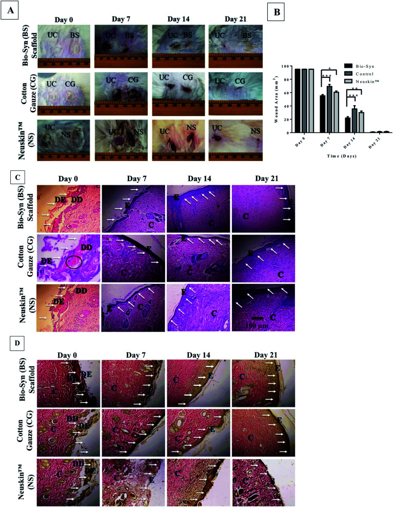
Fig. 6. (A) Representative photographs for 2nd degree burn wounds at days 0, 7, 14 and 21, (B) wound contraction area of wound tissue in the Bio-Syn, cotton gauze and Neuskin™ groups at days 0, 7, 14 and 21. The data are represented as the mean ± SD; n = 6 rats, (C) H&E stained histological section of wound tissue in the Bio-Syn, cotton gauze and Neuskin™ groups at days 0, 7, 14 and 21 (10× magnification), (D) Van Gieson’s (VG) stained histological section of wound tissue in the Bio-Syn, cotton gauze and Neuskin™ groups at days 0, 7, 14 and 21. The collagen stain is bright red, while the cytoplasm, muscle and fibrin stain is yellow. The white arrows indicate the epidermal healing process, while the black circles indicate ruptured blood capillaries. Abbreviations used in these images: “UC” untreated control, “DE” dead epidermis, “DD” dead dermis, “E” epidermis and “C” collagen fiber.

Fig. 6