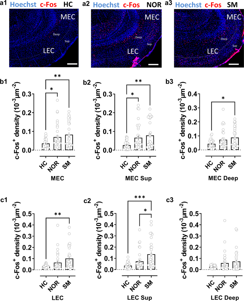
Figure 5. Social exploration preferentially activates lateral compared to medial entorhinal cortex.
a, Staining for c-Fos in lateral (LEC) and medial (MEC) entorinal cortices in horizontal brain slices from mice in control home-cage conditions (HC) or following novel object recognition (NOR) or social memory (SM) tasks. b, Quantification of c-Fos+ cell density in MEC in both deep and superficial layers combined (b1) or separated into superficial (b2) or deep layers (b3). Mice subjected to the NOR or SM task showed a significantly larger density of c-Fos+ cells than mice in HC conditions in both entire MEC and in superficial MEC layers. In deep MEC layers the increase over HC c-Fos+ staining was only significant following the SM task, although there was a trend in the NOR task. There was no significant difference in c-Fos+ density following SM compared to NOR task in total MEC (b1) or individual layers (b2, b3) c, In superficial and deep LEC layers combined (c1) and superficial LEC layers alone (c2), we observed a significant increase in c-Fos+ density compared to HC levels following the SM task, with no significant increase following the NOR task (although there was a trend). We saw no significant change in either SM or NOR tasks in deep LEC alone (c3). c-Fos+ density in superficial LEC was significantly greater following SM task compared to NOR task. HC: 27 sections from 4 animals, NOR: 32 sections from 4 animals; SM, 29 sections from 4 animals. Scale bar: 200 μm. *: p<0.05, **: p<0.01, ***: p<0.001 Holm-Sidak’s post hoc test after one-way ANOVA (in b1: F=5.238 p=0.0072; in b2: F=5.806 p=0.0043; in b3: F=4.217 p=0.0179; in c1: F=6.151 p=0.0032; in c2: F=9.399 p=0.0002; in c3: F=2.826 p=0.0648).

