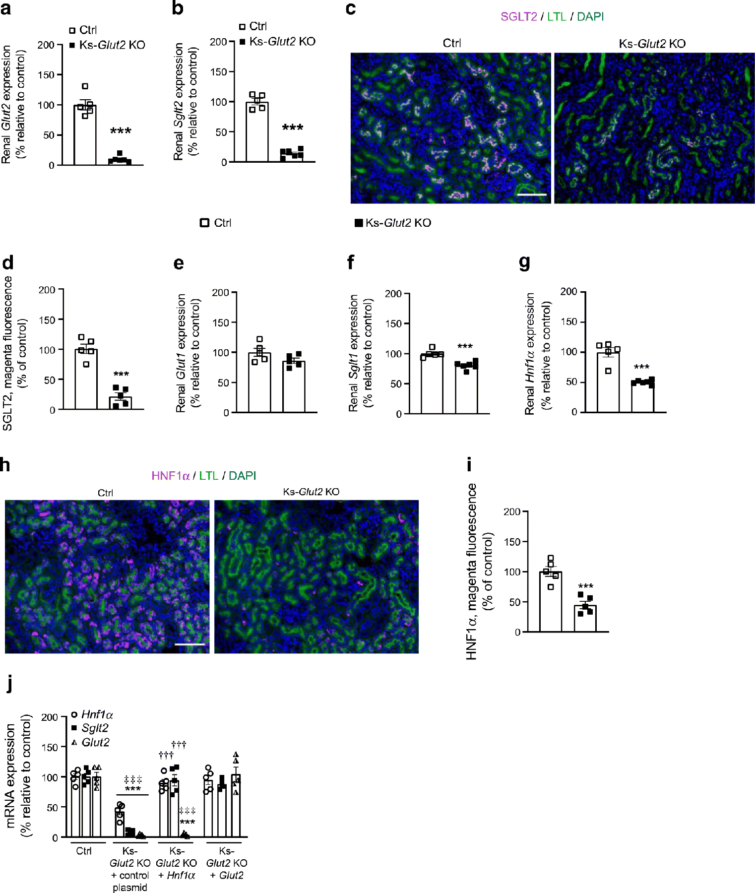
Fig. 6.

Long-term renal Glut2 deficiency almost abolishes the expression of Sglt2 by downregulating Hnf1α. (a, b) Expression of renal cortical Glut2 (a) and Sglt2 (b). (c, d) Immunofluorescence staining of renal SGLT2, with its quantification. Scale bar, 100 μm. (e–i) Expression of renal cortical Glut1 (e), Sglt1 (f) and Hnf1α (g), and immunofluorescence staining of renal HNF1α as well as its quantification (h, i), on the 21st day following renal Glut2 deficiency in 8- to 12-week-old male mice. Scale bar, 100 μm. (j) mRNA levels of Hnf1α, Sglt2 and Glut2 in primary renal proximal tubular epithelial cells isolated from 8- to 12-week-old male control (Glut2loxP/loxP) mice, or mice with kidney-specific loss of Glut2 (Ks-Glut2 KO) on the 21st day following renal Glut2 deficiency. Data are shown as means ± SEM, n=5 or 6. ***p<0.001 vs Ctrl; ‡‡‡p<0.001 vs Ks-Glut2 KO + Glut2; †††p<0.001 vs Ks-Glut2 KO + control plasmid group (two-tailed unpaired Student’s t test or two-way ANOVA followed by Bonferroni’s multiple comparison test). Ctrl, control Glut2loxP/loxP group; HNF1α, hepatocyte nuclear factor-1α; Ks-Glut2 KO, Glut2 knocked out specifically in the kidneys; Ks-Glut2 KO + Hnf1α or Glut2, primary renal proximal tubular epithelial cells isolated from Ks-Glut2 KO mice and transfected with Hnf1α or Glut2 expressing plasmids; LTL, Lotus tetragonolobus lectin, a marker of renal proximal tubules
