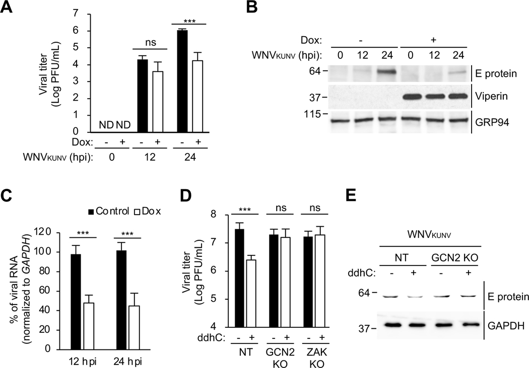
Figure 7. GCN2 and ZAK are required for the inhibition of viral protein synthesis and viral replication by viperin.
(A) 293T.iVip cells were treated with Dox and infected with West Nile virus Kunjin strain (WNVKUNV) at an MOI of 1. Viral titers were determined by plaque assay. PFU, plaque-forming unit. hpi, hours post-infection. ND, not detected.
(B)Immunoblot analysis of WNVKUNV E proteins in 293T.iVip cells. 293T.iVip cells were treated with Dox for 24 h, infected with WNVKUNV at an MOI of 1. Cell lysates were analyzed by immunoblotting with anti-flavivirus envelope (E) protein, anti-viperin and anti-GRP94 antibodies. hpi, hours post-infection.
(C) 293T.iVip cells were treated with Dox and then infected with WNVKUNV at an MOI of 1 for 24 h. Cells were harvested for RNA extraction. Viral RNA was determined by qRT-PCR and normalized to GAPDH.
(D-E) NT, GCN2 KO and ZAK KO cells were treated with ddhC for 24 h and infected with WNVKUNV at an MOI of 1 for 24 h. Viral titers were determined by plaque assay (D). PFU, plaque-forming unit. Cell lysates were analyzed by immunoblotting with anti-anti-flavivirus E protein and anti-GAPDH antibodies.
For (A), (C) and (D), data are shown as mean ± SD of three biological repeats (n = 3). ***p < 0.001 by unpaired Student’s t test. ns, not significant.

