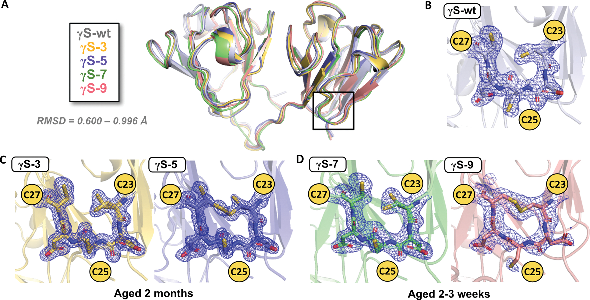
Figure 4, related to Figure S3 and S7. Crystal structures and electron density maps of the cysteine loop for aged γS wild-type and each variant.

(A) Alignment of the structures of γS-wt (PDB: 7N36, gray), γS-3 (PDB: 7N37, yellow), γS-5 (PDB: 7N38, blue), γS-7 (PDB: 7N39, green), and γS-9B (PDB: 7N3B, pink). All structures have Cα RMSDs less than 1 Å compared to γS-wt, PDB: 7N36. Magnified views of the cysteine loop region with 2FO-FC (contoured at 1σ) electron density maps for (B) γS-wt aged 1.5 months, (C) γS-3 and γS-5 aged 2 months and (D) γS-7 and γS-9B aged 2–3 weeks.
