
Figure 1.
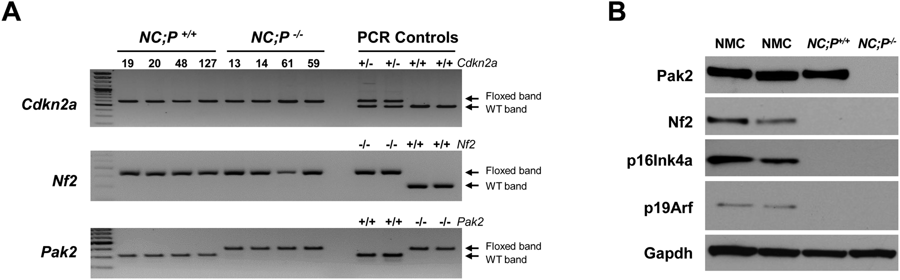
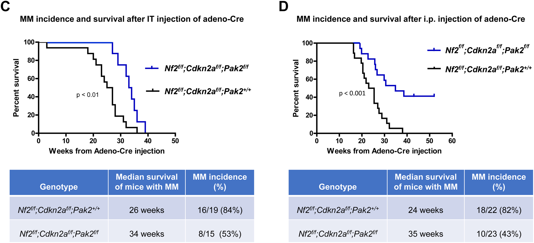
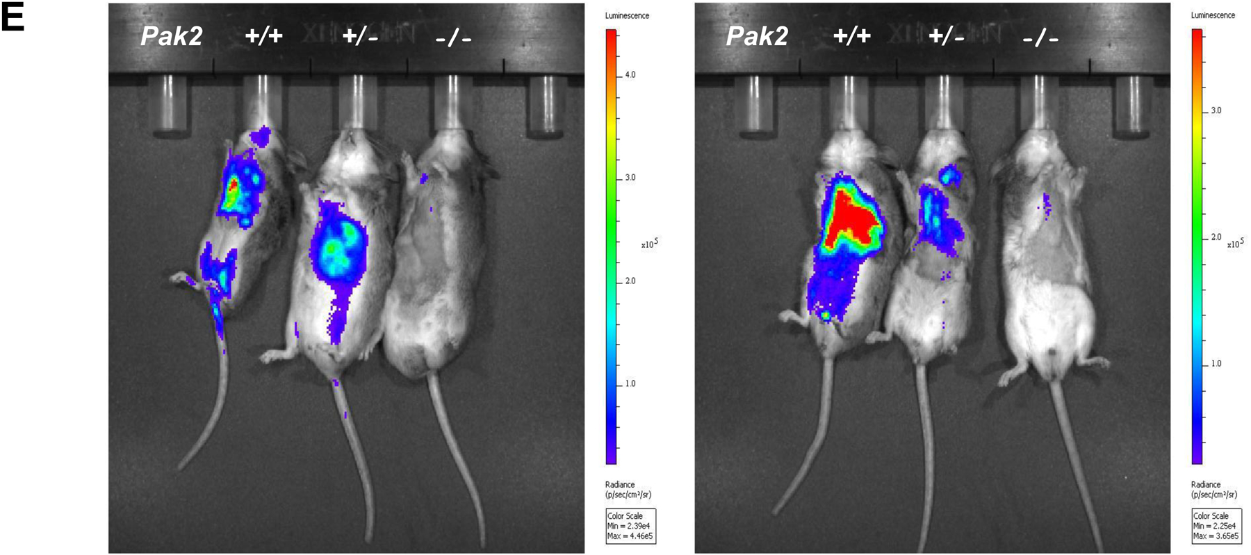
Genotyping and characterization of mesothelioma development in, Nf2f/f;Cdknaf/f;Pak2+/+ and Nf2f/f;Cdknaf/f;Pak2f/f mice injected intrathoracically or intraperitoneally with adeno-Cre virus. (A) Genotyping of tail DNA from four representative Nf2f/f;Cdknaf/f;Pak2+/+ mice and four Nf2f/f;Cdknaf/f;Pak2f/f mice that developed peritoneal mesothelioma. PCR controls for floxed and wild type alleles of Cdkn2a, Nf2 and Pak2 were from tail DNA of heterozygous and homozygous conditional knockout mice. (B) Immunoblotting of two early passage cell lines derived from NC;P−/− and NC;P+/+ peritoneal mesotheliomas demonstrating loss of expression of conditionally knocked out genes in tumor cells. NMC, normal mesothelial cells. (C, D) Malignant mesothelioma progression, incidence and Kaplan-Meier survival curves of cohorts of Nf2f/f;Cdknaf/f;Pak2+/+ and Nf2f/f;Cdknaf/f;Pak2f/f mice injected intrathoracically (C) or intraperitoneally (D) with adeno-Cre virus and succumbing to mesothelioma. Abbreviations: IT, intrathoracic (injection); i.p., intraperitoneal (injection); MM, malignant mesothelioma. (E) Bioluminescent imaging reveals delayed mesothelioma progression in mice with Nf2-null mesothelial lining and excision of one or both alleles of Pak2. ffLucR;Nf2f/f;Cdkn2af/f mice were crossed to Pak2f/f mice to generate offspring having wild type (+/+) Pak2 or with one or both floxed (+/f or f/f, respectively) Pak2 alleles. Mice were injected IT with adeno-Cre virus to excise floxed alleles of thoracic mesothelial lining cells. Infection with adeno-Cre virus also removes a floxed polyadenylation sequence before the ORF of a luciferase reporter transgene (LucR). The latter permits luciferase expression to monitor tumor progression, using D-luciferin as a substrate and bioluminescent imaging with an IVIS Imaging System. Shown is bioluminescent imaging on two sets of ffLucR;NC littermates with three different Pak2 genotypes. Mice were injected with D-luciferin 6 months (left panel) or 7 months (right) after IT injection of adeno-Cre virus; mice with excision of Pak2 show delayed tumor progression as indicated by reduced intensity of luminescent signals. Experiment was repeated four times with similar results.