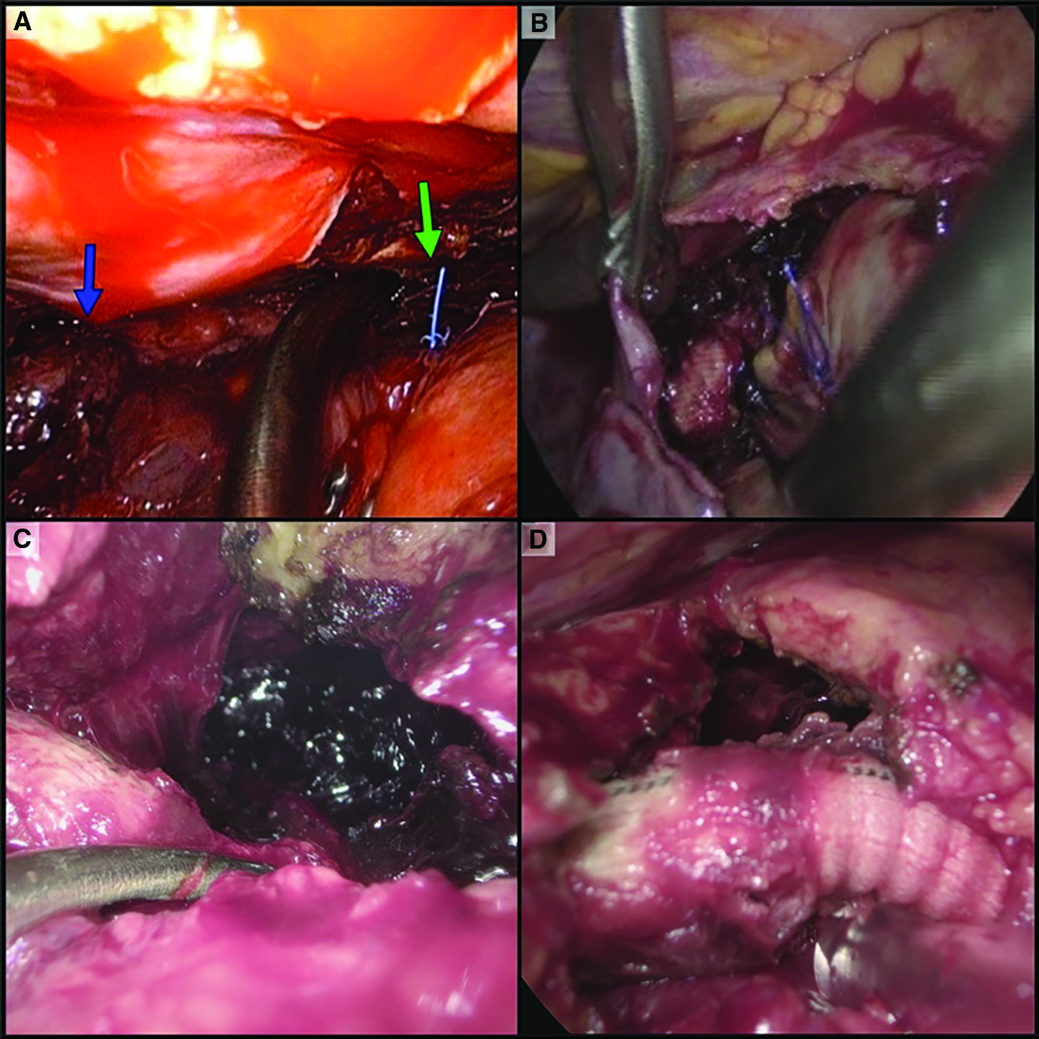
Fig. 2. Intraoperative view. (A) Diaphragm is on the left side, blue arrow indicating clotted blood, and green arrow indicating pacemaker wires. (B) Clots, fibrin, and empyema are visible in the pleural cavity surrounding the open pericardium. Hemopericardium evacuation was performed from the right side; threads of atrial cannulation are visible in the center. (C) Hemopericardium evacuation after LVAD implantation using VATS from the left side. Opened pericardium at the upper right corner, and clotted blood in the center. (D) Result of hemopericardium evacuation. Opened pericardium on the right side, and outflow graft (from left ventricle to aorta) in the center.

