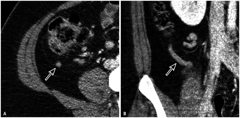
Fig. 2. A 21-year-old female with right lower quadrant pain.
A, B. Contrast-enhanced transverse (A) and coronal (B) low-dose CT images clearly show the normal appendix (arrows) in the abundant periappendiceal fat. The effective dose of the CT scan was 3 mSv, which was adjusted to the body size (body-mass index, 33.5 kg/m2) through automatic exposure control.

