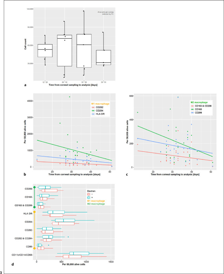
Fig. 3.
Macrophages in organ-cultured human corneas. a The total cell count per cornea varied over a wide range and decreased with increasing time in the organ culture (n = 24). To assess whether the macrophage populations decrease over time, the number of macrophages per 50,000 alive cells was analyzed at different time points. Corneas with different tissue culture times were digested and examined for cell marker expression (n = 24). Both, cells expressing pro-inflammatory (b) and regulatory markers (c) decreased with increasing time in organ culture. d To assess whether the cell culture conditions affected macrophages in human corneas, media with and without dextran were compared. Cornea halves were kept in the 2 different media for 24 h, followed by tissue processing and FC analysis (n = 10 per group). The presence or absence of dextran in tissue organ culture medium did not significantly alter the marker expression patterns (d). FC, flow cytometry.

