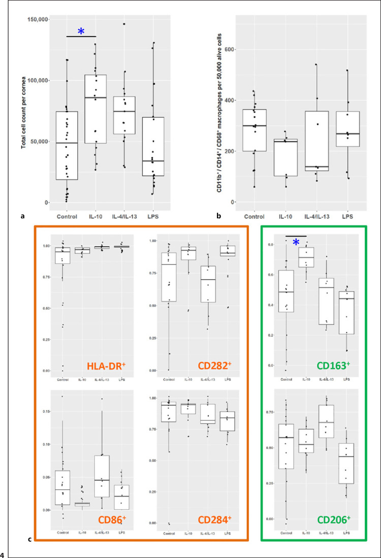
Fig. 4.
Polarization of human corneal macrophages especially monocyte-derived cells. a Corneal cell counts after the respective treatment (each point represents an individual cornea) showed statistically significant increased cell counts for corneas treated with IL-10 (one-way ANOVA p < 0.05). For analyzing macrophages, the cell marker expression was normalized to 50,000 live cells. b Macrophage cell count showed no statistical differences among the different groups. c The percentage of CD45+/CD11b+/CD14+/CD68+ macrophages after treatment revealed an altered macrophage phenotype for CD163+ cells after treatment with IL-10 (*p < 0.05, one-way ANOVA).

