
Fig. 5. LRP4 is the target of Ouabain and Digoxin.
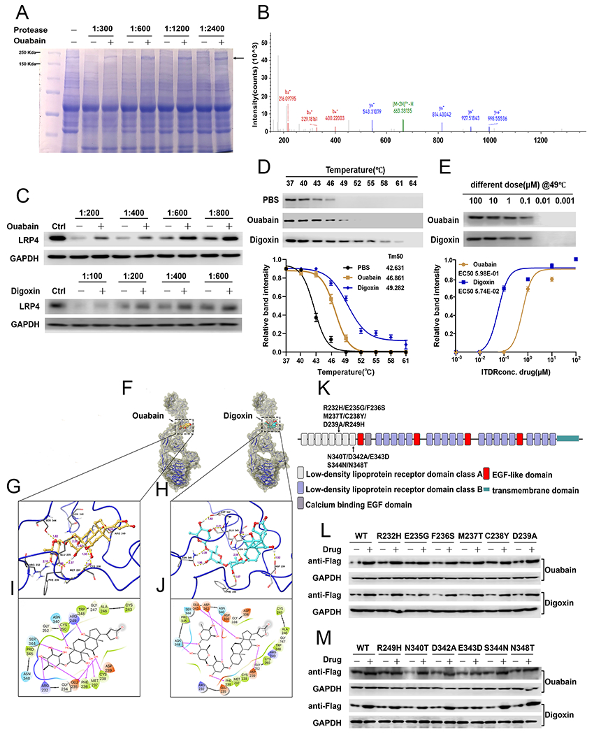
(A) Coomassie blue staining of DARTS assay. The band with molecular weight around 200 kDa was protected by Ouabain. (B) LRP4 adapted image from mass spectrometry. (C) C28I2 cells were digested with several dosages of protease with or without various concentrations of drugs, as indicated, then the level of LRP4 was assayed using Western blot. (D) C28I2 cell lysate was denatured under various temperatures and the protein level of LRP4 in control, Ouabain-treated and Digoxin-treated groups were assayed using Western blot and densitometry analysis curve. (E) Isothermal dose response with serial concentrations of drug. Protein level of LRP4 was measured via Western blot with associated curve. (F) Overview of IFD-predicted binding positions of Ouabain and Digoxin in LRP4 aa 146-737 monomer. LRP4 is shown by ribbons along with yellow surface of 70% transparency. Ouabain and Digoxin are shown by CPK representation with the following color scheme: carbon-faded orange (Ouabain) or teal (Digoxin), oxygen-red, polar hydrogen-white. Non-polar hydrogen atoms are not shown. (G, H) IFD-predicted docked LRP4-Ouabain complex (G) and LRP4-Digoxin complex (H) with the ligand depicted in ball and stick and the important interacting residues depicted as sticks. Hydrogen bonds are represented by dotted yellow lines and the distance of hydrogen bonds are measured in Å. (I, J) The 2D interaction diagrams of Ouabain (I) and Digoxin (J) docked with LRP4 aa 146-737. The amino acids within 4 Å to the ligand are shown as colored bubbles, where polar residues are cyan, hydrophobic residues are green, positively charged residues are purple, and negatively charged residues are red. Hydrogen bonds are shown by magenta arrows. (K) Schematic view of LRP4 receptor domain organizations and localization of mutations tested. (L, M) DARTS assay for serial point mutants of LRP4. C28I2 cells were transfected with the plasmid expressing various Flag-tagged LRP4 mutants, as indicated. Mutants of LRP4 were detected by Flag antibody. The values are mean±SEM of at least 3 independent experiments.